Ultrasound diagnostic apparatus

A diagnostic device, ultrasonic technology, applied in the direction of acoustic wave diagnosis, infrasonic wave diagnosis, ultrasonic/sonic wave/infrasonic wave diagnosis, etc., which can solve problems such as complex structure of the device

Active Publication Date: 2009-03-25
OLYMPUS CORP
View PDF3 Cites 7 Cited by
  • Summary
  • Abstract
  • Description
  • Claims
  • Application Information

AI Technical Summary

Problems solved by technology

[0006] The biplanar ultrasonic diagnostic devices using two transducer groups disclosed in Japanese Patent Application Laid-Open No. 8-56948 and Japanese Patent Laid-Open No. 2004-135693 require separate sound ray synthesis tables and transmission and reception tables corresponding to each transducer group. part and image generation part, etc., the device structure becomes complicated
[0007] In addition, in the ultrasonic diagnostic apparatus disclosed in Japanese Patent Laid-Open No. 2002-177278, only a probe using a plurality of vibrator groups having the same shape and driving method is disclosed, and the same control device, etc. are used for switching.

Method used


Image

  • Ultrasound diagnostic apparatus
  • Ultrasound diagnostic apparatus
  • Ultrasound diagnostic apparatus

Examples


Embodiment Construction

[0032] Hereinafter, embodiments of the present invention will be described with reference to the drawings.

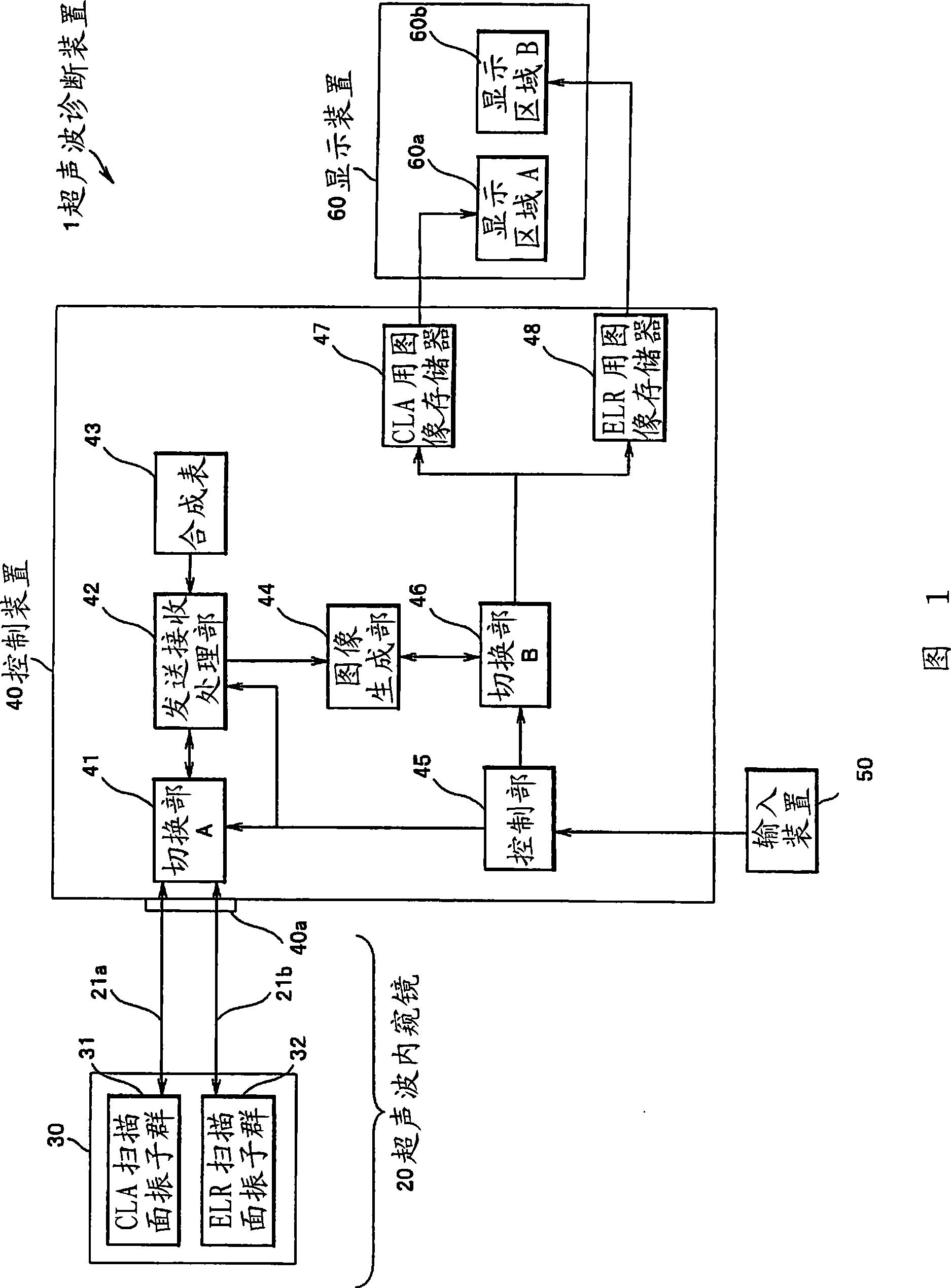
[0033] FIG. 1 is a configuration diagram of an ultrasonic diagnostic apparatus 1 according to an embodiment of the present invention. The ultrasonic diagnostic apparatus 1 of the present embodiment includes: an ultrasonic endoscope 20 , a control device 40 , an input device 50 connected to the control device 40 to operate the control device 40 , and a control device connected to the control device 40 to display 40 to the display device 60 of the video signal obtained.

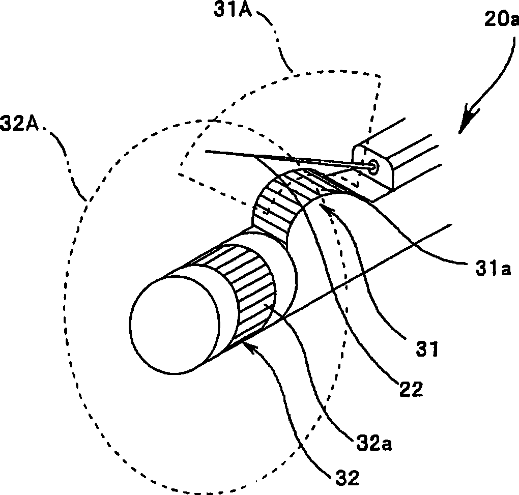
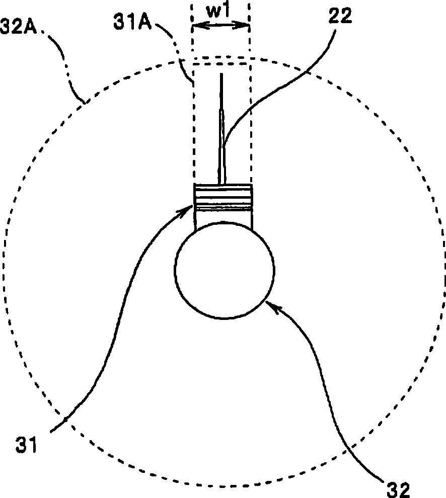
[0034] The ultrasonic endoscope 20 is inserted into a body cavity or the like, transmits an ultrasonic beam to an observation target site, receives reflected waves reflected from the acoustic impedance boundary of the observation target site, and obtains echo signals. The control device 40 is connected to the ultrasonic endoscope 20 with the cables 21a and 21b through the connector 40a, controls the sen...


PUM


Abstract

An ultrasound diagnostic apparatus (1) having a plurality of transducer groups (31 and 32) which are provided at a distal end portion of a probe (20a), and are configured by a plurality of transducers arranged in a circumferential form or an arc form having a same radius of curvature, and a common sound ray synthesis table (43) storing control data of the respective transducer groups (31 and 32).

Description

technical field [0001] The present invention relates to an ultrasonic diagnostic apparatus, and more particularly to an ultrasonic diagnostic apparatus provided with an ultrasonic endoscope having a plurality of transducer groups scanning in mutually orthogonal planes at the distal end of a probe. Background technique [0002] In recent years, a method of inserting an ultrasonic endoscope into a living body, discovering a lesion in the body from its optical image, irradiating ultrasonic waves, and diagnosing ultrasonic tomographic images of the lesion from the reflected waves has become widespread. In addition, the following method is also implemented, using a puncture needle under the guidance of an optical image / ultrasound tomographic image, visually identifying and performing puncture to absorb cells, and confirming the diagnosis by absorbing cells. [0003] When making such a diagnosis, it is necessary to collect the tissue accurately, and as a collection method, there a...

Claims


Application Information

Patent Timeline
Patent Type & Authority Applications(China)
IPC IPC(8): A61B8/12A61B8/14
CPCA61B8/461A61B8/145A61B8/4488A61B8/463G01S15/8929G01S15/892G01S7/52074A61B8/0833A61B8/12A61B8/0841A61B8/445A61B8/00A61B1/00
Inventor 日比靖
Owner OLYMPUS CORP
Why Eureka
Try Eureka