Nematophagous fungi infectious extracellular serine proteinase crystal morphology

A technology of serine protease and nematophagous fungi, which is applied in the fields of molecular biology and applied microbiology, and can solve the problems of undiscovered public reports and the like

Inactive Publication Date: 2009-05-20
YUNNAN UNIV
View PDF0 Cites 5 Cited by
  • Summary
  • Abstract
  • Description
  • Claims
  • Application Information

AI Technical Summary

Problems solved by technology

[0006] Through document search, do not find the public report of the same document as the content of the present invention

Method used


Image

  • Nematophagous fungi infectious extracellular serine proteinase crystal morphology
  • Nematophagous fungi infectious extracellular serine proteinase crystal morphology
  • Nematophagous fungi infectious extracellular serine proteinase crystal morphology

Examples


Embodiment 1

[0035] Example 1: Structural analysis of the extracellular serine protease from Verticillium lanceolata Lecanicillium psalliotae

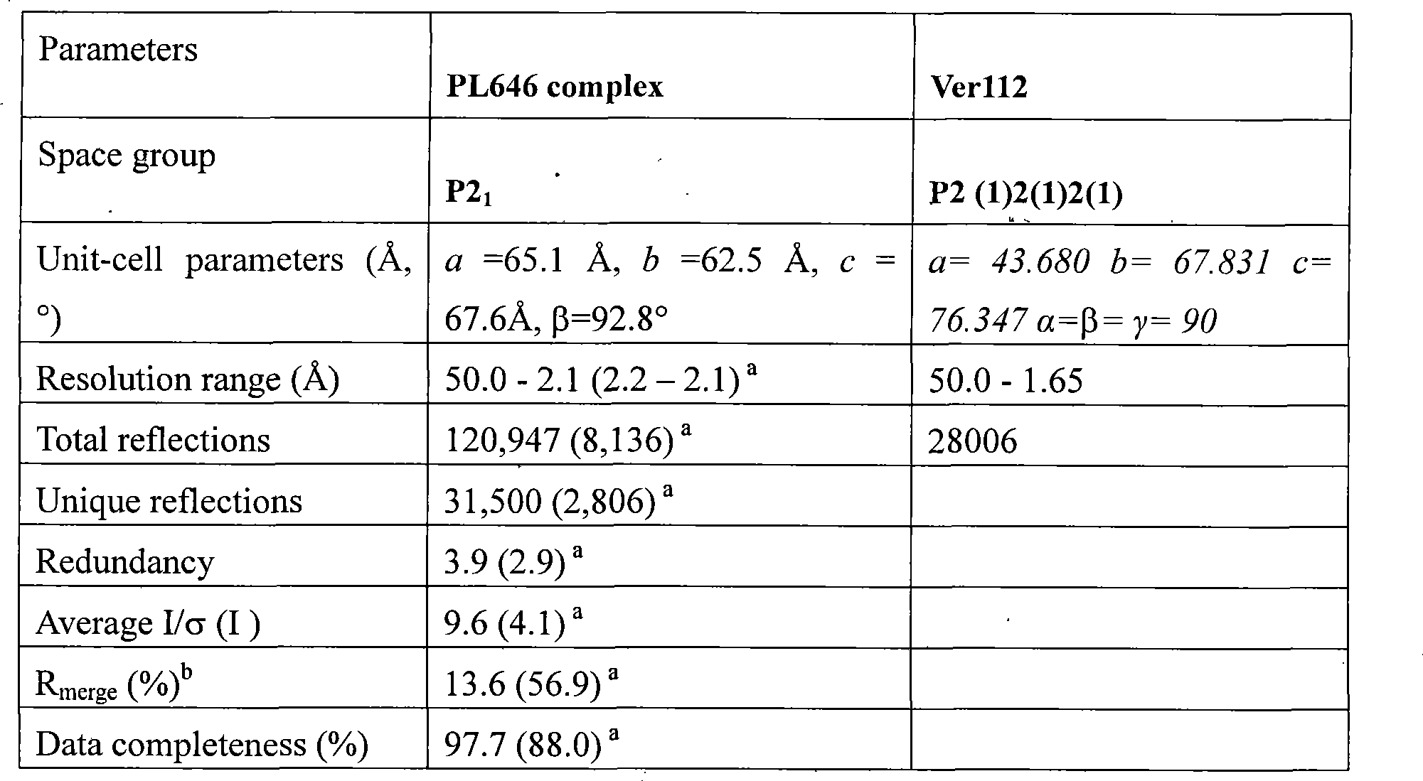
[0036] Inoculate the strain of Verticillium psalliotae (Lecanicillium psalliotae, YMF1.00112) with the preservation number CGMCC No. 1312 cultured on the PDA plate in the enzyme production medium, and culture it on a shaker at 28° C. at 150 rpm for 6 days. The fermentation broth was filtered and the supernatant was precipitated with ammonium sulfate. Fractions having protease activity were dissolved in 10 mM PBS (pH 6.0) buffer and purified by cation exchange chromatography. The enzymatically active eluate was further purified by molecular sieves. The electrophoretic pure protease Ver112 was obtained, concentrated and then crystallized to obtain protease crystals. The three-dimensional structure of the protease was obtained by X-ray diffraction of the crystal and processed with software such as HKL2000 and ccp4.

Embodiment 2

[0037] Example 2: Structural Analysis of Paecilomyces lilacinus Extracellular Serine Protease

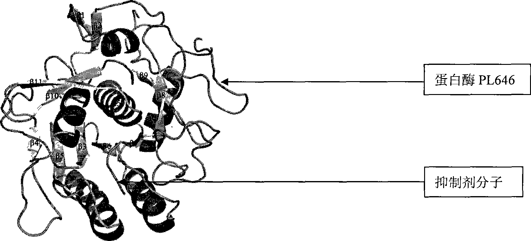
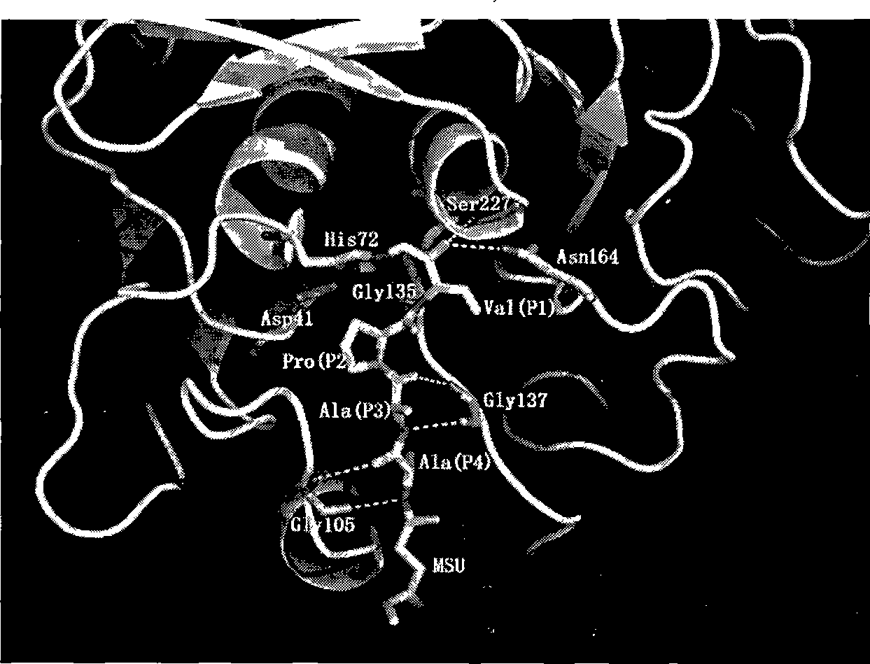
[0038] Paecilomyces lilacinus (M-14) cultured on the PDA plate, the preserved strain of CGMCC No0241. was inoculated in the enzyme production medium, and cultured on a shaker at 28° C. at 150 rpm for 6 days. The fermentation broth was filtered and the supernatant was precipitated with ammonium sulfate. The protease active fraction was dissolved in 10 mMPBS (pH 6.0) buffer and purified by cation exchange chromatography. The enzymatically active eluate was further purified by molecular sieves. The electrophoretic pure protease PL646 was obtained, concentrated and then crystallized to obtain protease crystals. The three-dimensional structure of the protease was obtained by X-ray diffraction of the crystal and processed with software such as HKL2000 and ccp4.

[0039] The crystal structure of the extracellular alkaline serine protease of the nematode fungus obtained by the above-ment...


PUM


Abstract

The invention relates to an extracellular serine protease crystal structure with nematophagous fungi infectivity and belongs to the fields of molecular biology and applied microbiology. The invention implements liquid cultivation with Lecanicillium psalliotae YMF1.00112 and Paecilomyces lilacinus M-14, fractional precipitation of ammonium sulphate with a fermented liquid, and dissolution of protease-containing active component with buffer solution, and then obtains raw enzyme solution. Cation exchange chromatography and molecular sieve purification of the raw enzyme solution are implemented. After purified enzyme is obtained, concentration processing is implemented so as to obtain an enzyme solution with higher concentration. Hanging drop vapor diffused crystallization of the enzyme solution with higher concentration is implemented. The crystallization conditions are filtered with a reagent kit. Enough diffraction pictures of obtained crystal are collected through diffraction on an X-ray machine. The diffraction pictures are processed with HKL2000, protein structure is analyzed and optimized with software O, ccp4 and the like. The invention discloses the extracellular serine protease crystal structure for the first time and provides foundation for improving the hydrolysis activity of protease and the infectivity of nematophagous fungi.

Description

Technical field: [0001] The invention relates to the crystal structure of nematode fungus infective extracellular serine protease determined by X-ray crystal diffraction method, and belongs to the fields of molecular biology and applied microbiology. Background technique: [0002] Nematodes are a class of linear multicellular protozoa widely distributed in seawater, freshwater and soil, some of which can parasitize plants or animals. Plant parasitic nematodes can cause serious diseases of a variety of commercial crops, resulting in huge economic losses. The most serious nematodes include root-knot nematode (Meloidogyne spp.), cyst nematode (Heterodera spp.) and pine wood nematode (Bursaphelenchus xylophilus). Although the application of chemical pesticides has a good control effect on pathogenic nematodes, chemical pesticides are likely to cause environmental pollution, pesticide residues, and nematodes are prone to drug resistance. Therefore, the research on nematicidal bi...

Claims


Application Information

Patent Timeline
Patent Type & Authority Applications(China)
IPC IPC(8): C12N9/58
Inventor 张克勤饶子和孟照辉娄智勇杨金奎梁连铭
Owner YUNNAN UNIV
Why Eureka
Try Eureka