Method for largely expanding late endothelial progenitor cells from peripheral blood

A technology of endothelial progenitor cells and peripheral blood, which is applied in the biological field, can solve the problems of fewer late endothelial progenitor cell colonies, affecting clinical efficacy, and hindering the prospect of clinical use.

Inactive Publication Date: 2011-07-27
吴立华
View PDF0 Cites 2 Cited by
  • Summary
  • Abstract
  • Description
  • Claims
  • Application Information

AI Technical Summary

Problems solved by technology

However, the current problem is that the content of endothelial progenitor cells in the peripheral blood of adults is scarce, especially those with cardiovascular risk factors. The elderly and heart failure patients who need EPC treatment have a relatively small number of EPCs in their bodies, and it is known from the analysis of existing clinical data In some disease patients, the function of EPCs to form blood vessels is reduced. In short, the rare and functionally reduced EPCs limit its effective clinical application. Therefore, obtaining high-quantity and high-quality EPCs is a difficulty that needs to be solved urgently in the field of EPCs research.
The biggest disadvantage of the traditional method is that there are fewer colonies of advanced endothelial progenitor cells, and only two colonies of late endothelial progenitor cells can be obtained per 100ml of normal human peripheral blood, so the cell acquisition rate is low and it takes a long time. The laboratory stage is a waste of peripheral blood required In terms of clinical application, the small amount of cells obtained affects its clinical efficacy and hinders its clinical application prospects

Method used

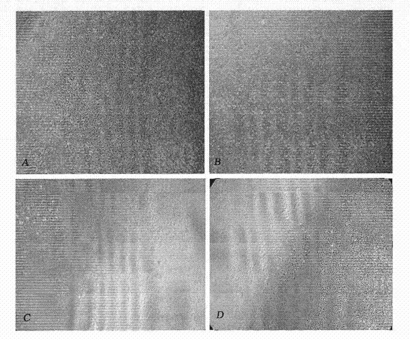
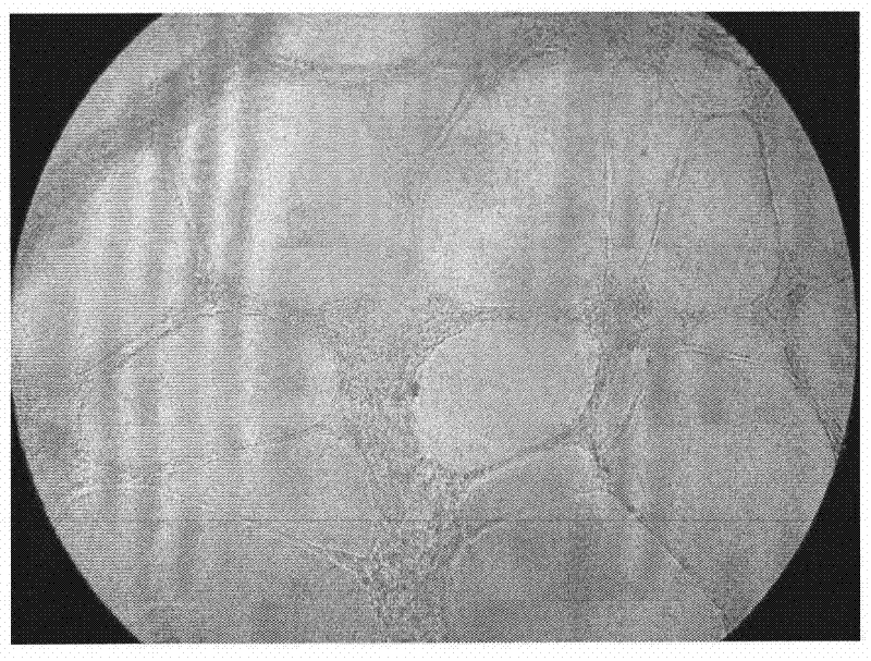
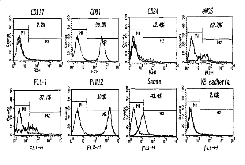

Image

  • Method for largely expanding late endothelial progenitor cells from peripheral blood
  • Method for largely expanding late endothelial progenitor cells from peripheral blood
  • Method for largely expanding late endothelial progenitor cells from peripheral blood

Examples


preparation example Construction

[0031] (2) Preparation of trophoblast cells in a culture dish: 1×10 late endothelial progenitor cells or HUVECs (umbilical cord endothelial cells) obtained from previous culture 3 Cells were planted in 1% collagen-coated 6-well plates, with EGM2 as the culture medium, at 37°C, 5% CO 2 Cultivate in the incubator for 6 hours, and after the cells adhere to the wall, they are irradiated with a dose of 10Gy to inhibit their growth.

[0032] (3) In vitro culture of mononuclear cells: the isolated mononuclear cells were planted into the prepared 6-well plate, with EGM2 as the culture medium, at 37 ° C, 5% CO 2 Cultured in an incubator, changing the medium every other day.

[0033] The above-mentioned in vitro culture medium is a uniformly sold culture medium on the market; mononuclear cells are divided into 5×10 7 Cells were seeded into 6-well plates coated with 1% collagen; the coating concentration of collagen was 5-10ul / cm 2 ; Trophoblast radiation dose 10Gy.

[0034] (4) Iden...

Embodiment 1

[0041] 1. Isolation of human peripheral blood mononuclear cells: Take 100ml of human venous anticoagulant blood, dilute it with PBS balanced salt solution 1:1, slowly spread 32ml of the diluted solution onto the 18ml of Ficoll separation solution along the tube wall, and use 2000r / m, centrifuged for 30 minutes, after density gradient centrifugation, suck out the white ring-shaped cloud layer, then wash twice with PBS balanced salt solution at 1000r / m, 10 minutes, and finally resuspend the cells with culture medium, and count with placenta blue cell.

[0042] 2. Preparation of trophoblast cells in a culture dish: 1×10 late endothelial progenitor cells or HUVECs obtained from previous culture 3 Cells were planted in 1% collagen-coated 6-well plates, with EGM2 as the culture medium, at 37°C, 5% CO 2 Cultivate in the incubator for 6 hours, and after the cells adhere to the wall, they are irradiated with a dose of 10Gy to inhibit their growth.

[0043] 3. In vitro culture of mon...


PUM


Abstract

The invention discloses a method for largely expanding endothelial outgrowth cells from peripheral blood. Mononuclear cells separated from the peripheral blood are planted to a pretreated culture dish spread with endothelial outgrowth cells or umbilical endothelial cells as nutritive layers to obtain more endothelial outgrowth cell colonies, and then the endothelial outgrowth cells are largely expanded. By previously spreading the nutritive layer cells containing endothelial cells in the culture dish before planting the mononuclear cells of the peripheral blood, and then planting the mononuclear cells in the culture dish, the method can obtain colony numbers of the endothelial outgrowth cells 20 times more than the traditional method about two weeks in advance, and can obtain the endothelial outgrowth cells with high quality, high quantity and treatment value from the limited peripheral blood.

Description

technical field [0001] The invention relates to the field of biotechnology, in particular to a culture method for obtaining more late endothelial progenitor cell colonies from human peripheral blood so as to amplify the late endothelial progenitor cells in large quantities. Background technique [0002] Asahara et al. reported in 1997 that adult peripheral blood CD34 + Cell populations can be induced to differentiate into endothelial-like cells in vitro. These cells express a series of endothelial cell antigens and have the function of participating in angiogenesis in vivo. They are named endothelial progenitor cells (EPCs), thus renewing angiogenesis The concept of angiogenesis exists not only in adults, but also in the adult body. With the in-depth study of endothelial progenitor cells, Hur J et al. reported that EPCs can be divided into early EPCs and late EPCs. As they described, a certain density of mononuclear cells derived from peripheral blood was seeded into fibron...

Claims


Application Information

Patent Timeline
Patent Type & Authority Patents(China)
IPC IPC(8): C12N5/071
Inventor 吴立华宋增璇韩忠朝李尚珠翟琼莉马凤霞
Owner 吴立华
Why Eureka
Try Eureka