Method for structural information fused electrical impedance tomography
A technology of electrical impedance tomography and structural information, applied in the direction of diagnosis, diagnostic recording/measurement, medical science, etc., to achieve the effect of improving image quality, improving analysis ability, and improving accuracy
- Summary
- Abstract
- Description
- Claims
- Application Information
AI Technical Summary
Problems solved by technology
Method used
Image
Examples
Embodiment 1
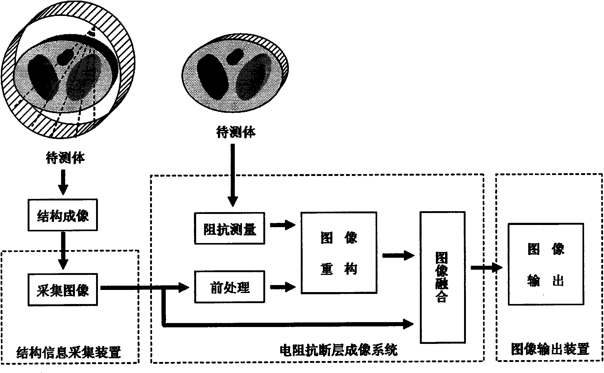
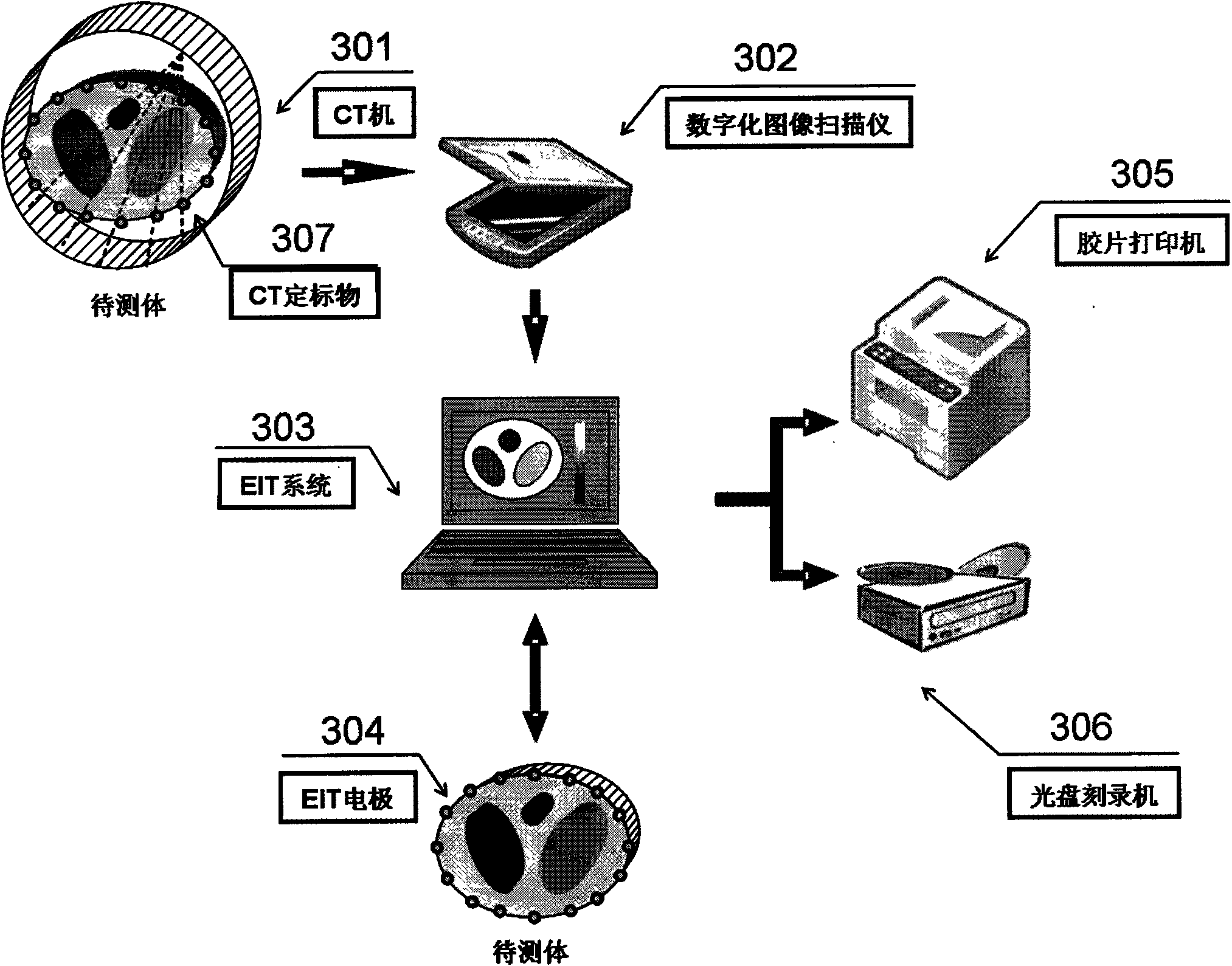
[0038] Embodiment 1, with reference to figure 1 and figure 2 The devices required in this embodiment include a CT machine 301 for detecting the object to be tested to realize structural imaging, that is, a structural information acquisition device to collect images, a digital imaging scanner 302, an electrical impedance tomography system (EIT) 303, and an electrical impedance tomography imaging system. Professional electrode and electrode belt 304, film printer 305, CD recorder 306 and CT calibration object 307.
[0039]In the embodiment of the present invention, the object to be tested is a cross-sectional area of the chest of a human body, and CT images with relatively high resolution are used for structural imaging. The structural information acquisition device is a digital image scanner capable of scanning CT slices, and the image output device is a film printer capable of printing CT slices.
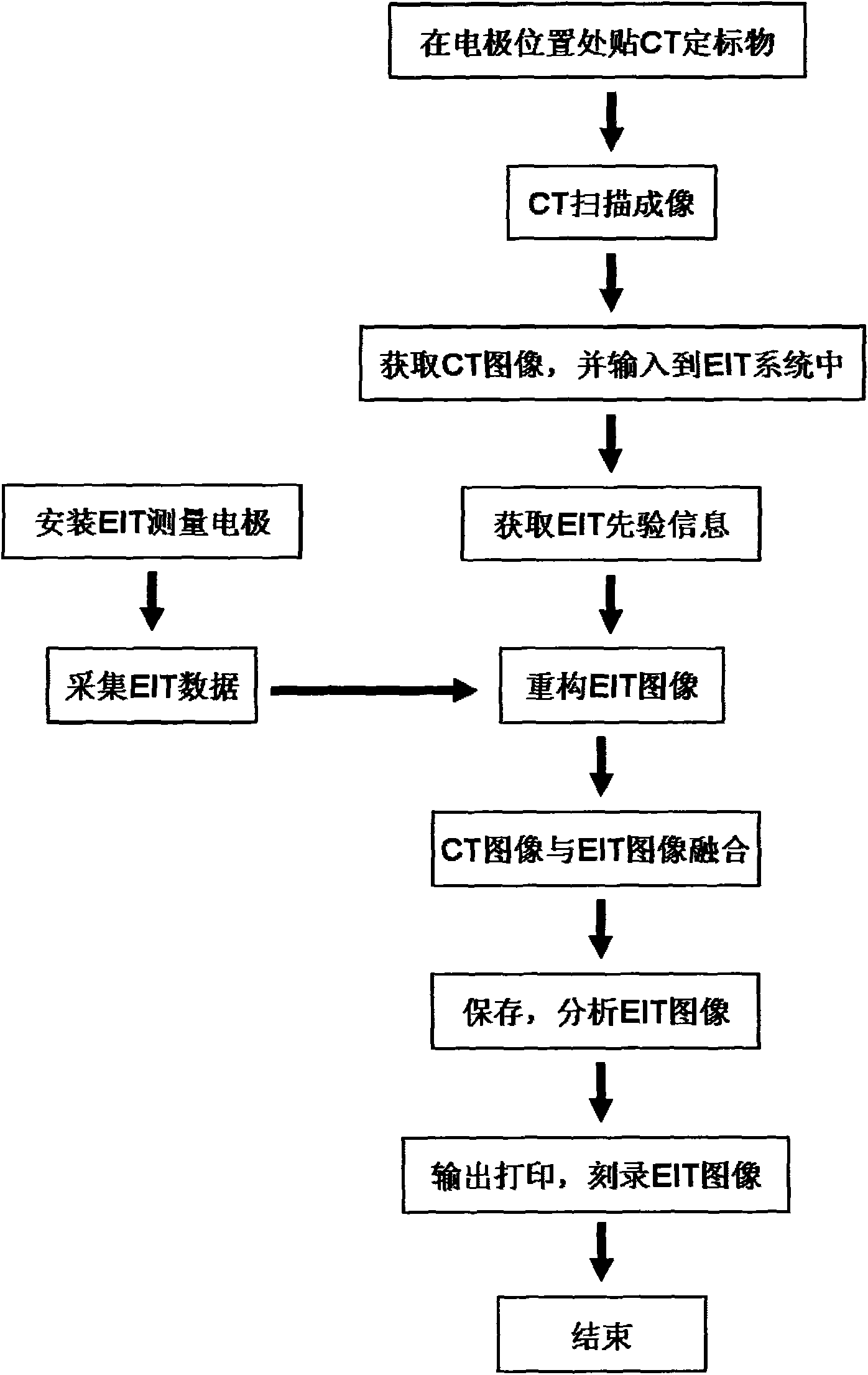
[0040] In the flow of the embodiment such as image 3 shown.
[0041] Fi...
Embodiment 2
[0042] Example 2: In this example, refer to figure 1 , Figure 4 As shown, in the embodiment of the present invention, the object to be measured is a cross-sectional area of the human head, and the structural image adopts a higher-resolution MRI image. The structural information collection device is a standard hospital information system network (Hospital Information Systems, HIS), and the image output device is a remote communication network. Its process is as follows Figure 4 shown. Use MRI to collect MRI images of the human head. The EIT system directly reads the MRI images through the HIS system and extracts the EIT prior information. Step is identical with embodiment 1.
Embodiment 3
[0043] Example 3: In this example, refer to figure 1 , figure 2 and image 3 As shown, in the embodiment of the present invention, instead of pasting CT calibration objects, optical or electromagnetic six-dimensional position sensors are used to directly obtain the position information of the pasted electrodes, and input them into the EIT system for use as prior information. All the other steps are the same as in Example 1.
[0044] In the following, the common processing methods in each preferred embodiment of the present invention will be further described in detail.
[0045] Pretreatment method
[0046] The EIT preprocessing method described in the present invention refers to the method of extracting EIT prior information from the structural image, and the EIT prior information includes but not limited to the boundary information of the object to be tested, the position information of the electrode pasting place, the boundary information of the internal structure of the...
PUM
 Abstract
Description
Claims
Application Information
 - R&D
- Intellectual Property
- Life Sciences
- Materials
- Tech Scout
- Unparalleled Data Quality
- Higher Quality Content
- 60% Fewer Hallucinations
Browse by: Latest US Patents, China's latest patents, Technical Efficacy Thesaurus, Application Domain, Technology Topic, Popular Technical Reports.
© 2025 PatSnap. All rights reserved.Legal|Privacy policy|Modern Slavery Act Transparency Statement|Sitemap|About US| Contact US: help@patsnap.com


