Optimized quantitative method of blood volume in magnetic resonance perfusion imaged image

A technology of perfusion imaging and quantification method, which is applied in the directions of using nuclear magnetic resonance imaging system for measurement, magnetic resonance measurement, and magnetic variable measurement, etc. The effect of suppressing quantification error and blood volume quantification error

Inactive Publication Date: 2010-06-02
NORTHWESTERN POLYTECHNICAL UNIV
View PDF1 Cites 13 Cited by
  • Summary
  • Abstract
  • Description
  • Claims
  • Application Information

AI Technical Summary

Problems solved by technology

The second and subsequent multiple passes will introduce significant overestimation error
The disadvantage

Method used


Image

  • Optimized quantitative method of blood volume in magnetic resonance perfusion imaged image
  • Optimized quantitative method of blood volume in magnetic resonance perfusion imaged image
  • Optimized quantitative method of blood volume in magnetic resonance perfusion imaged image

Examples


Embodiment Construction

[0027] Now in conjunction with embodiment, accompanying drawing, the present invention will be further described:

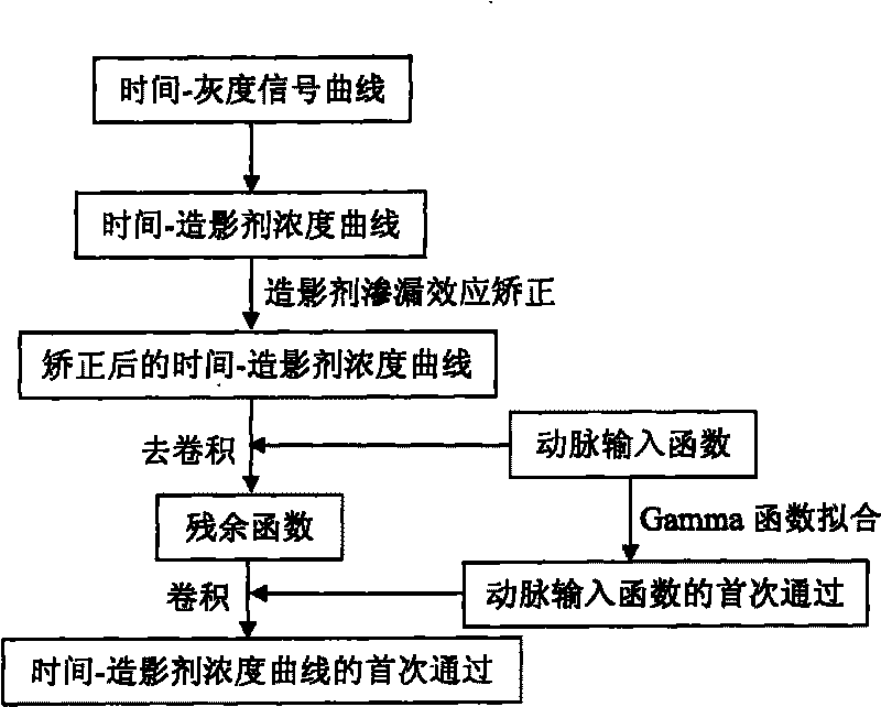
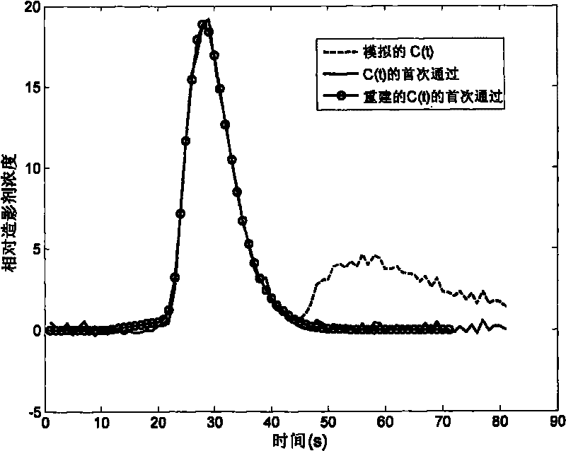
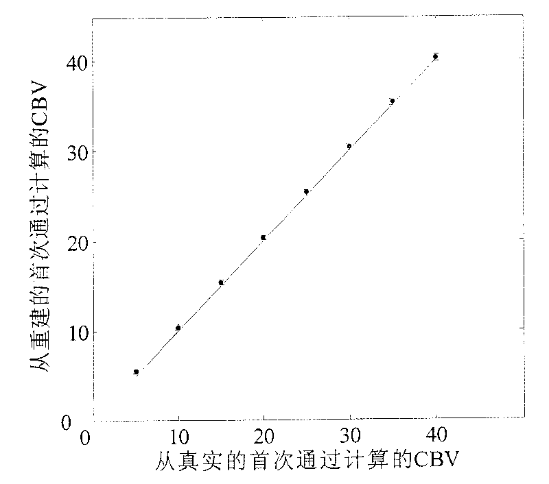
[0028] In this embodiment: firstly, the time-grayscale curve of the three-dimensional pixel in the perfusion imaging is converted into a time-contrast agent concentration curve. Then, the contrast agent leakage effect in the time-contrast agent concentration curve is removed by a contrast agent leakage correction algorithm. Secondly, select an arterial input function with a typical double-peak shape in the vicinity of the intracranial main central artery, input the obtained arterial input function into the function, and perform a deconvolution operation on the time-contrast agent concentration curve obtained in the previous step to obtain an approximate A residual function with a unimodal shape. Thirdly, use the gamma function to fit the selected arterial input function to obtain its first-pass component. Finally, the first pass of the arterial input function i...


PUM


Abstract

The invention relates to an optimized quantitative method of blood volume in a magnetic resonance perfusion imaged image. The optimized quantitative method is technically characterized by comprising the steps of: selecting an artery input function with a typical double-peak shape from an encephalic main central artery of the magnetic resonance perfusion imaged image; carrying out a convolution operation on a contrast agent concentration time sequence signal and the artery input function to obtain a residual function with similar single-peak shape; using gamma function fitting to select the artery input function to obtain a first pass of the artery input function; carrying out the convolution operation on the first pass of the artery input function which is obtained by fitting and the residual function, thus reestablishing a first pass component in an initial time-contrast agent concentration curve; and quantizing the blood volume by using the reestablished first pass component and inhibiting quantization error. By combining a first pass reestablishing algorithm with a contrast agent percolation effect correction algorithm, the invention furthers to inhibit the blood volume quantization error.

Description

technical field [0001] The invention relates to an optimized method for quantifying blood volume in magnetic resonance perfusion imaging images, which is a method for reducing quantification errors caused by contrast agent leakage effect and blood circulation in quantifying blood volume by using perfusion nuclear magnetic resonance imaging . It belongs to the medical image processing of blood volume in magnetic resonance perfusion imaging. Background technique [0002] Modern medical imaging is not satisfied with the study of morphological changes of lesions, but has developed to reflect the physiological and pathological changes of tissues and organs, and even functional imaging. Brain magnetic resonance perfusion imaging is one of the methods of brain functional imaging. It obtains microcirculation information by studying the blood perfusion state of brain tissue and the degree of tissue vascularization, so as to reveal the biological behavior and prognosis of tumors. Th...

Claims


Application Information

Patent Timeline
IPC IPC(8): G01R33/56
Inventor 郭雷胡新韬张拓聂晶鑫李刚刘天明李凯明
Owner NORTHWESTERN POLYTECHNICAL UNIV
Why Eureka
Try Eureka