Method for rapidly detecting cow and sheep derived components

A sheep source, cattle and sheep technology, applied in the field of molecular biology, to achieve the effect of easy operation

Inactive Publication Date: 2010-06-09
CENT LAB TIANJIN ACADEMY OF AGRI SCI
View PDF0 Cites 3 Cited by
  • Summary
  • Abstract
  • Description
  • Claims
  • Application Information

AI Technical Summary

Problems solved by technology

However, the bovine forward outer primer COW-F3, the bovine reverse outer primer COW-B3, the bovine forward inner primer COW-FIP, the bovine reverse inner primer COW-BIP; the sheep forward outer primer SHEEP- F3, the reverse outer primer SHEEP-B3 of sheep origin, the forward inner primer SHEEP-FIP of sheep origin, and the reverse inner primer SHEEP-BIP of sheep origin The method of constant temperature amplification to detect ingredients of cattle and sheep origin has not been reported in the literature

Method used


Image

  • Method for rapidly detecting cow and sheep derived components
  • Method for rapidly detecting cow and sheep derived components
  • Method for rapidly detecting cow and sheep derived components

Examples


Embodiment 1

[0049] (1) Reagent: Bst DNA polymerase large fragment produced by BioLabs (NEW ENGLAND) and 10 times Buffer solution; specific primer mixture; 4M betaine solution; 0.2M MgSO 4 solution;

[0050] (2) Amplification reaction system: the total volume of the amplification reaction is 25 μL, and its various components are: 10×Buffer 2.5 μL, 4M betaine 6.25 μL, 0.2M MgSO 4 0.25 μL, 1 μL of mixed primers, 3.5 μL of 10 μM dNTPs, 1 μL of 8000 U / L Bst DNA polymerase large fragment, 1 μL of template DNA, make up to 25 μL with sterilized deionized water, mix well and centrifuge;

[0051] (3) Amplification reaction procedure: proceed at 63°C for 60 minutes, and keep at 80°C for 2 minutes, store at 4°C;

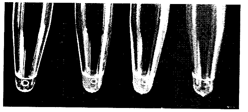
[0052] (4) After the amplification reaction, 15 μL of the system solution was taken, and 1 μL of 1000×SYBRGreen intercalator was directly added to the amplification tube, shaken and mixed, and the results were observed with the naked eye. Tubes without amplification reactions will be ora...

Embodiment 2

[0054] (1) Reagent: Bst DNA polymerase large fragment produced by BioLabs (NEW ENGLAND) and 10 times Buffer solution; specific primer mixture; 4M betaine solution; 0.2M MgSO 4 solution;

[0055] (2) Amplification reaction system: the total volume of the amplification reaction is 25 μL, and its various components are: 10×Buffer 2.5 μL, 4M betaine 6.25 μL, 0.2M MgSO 4 , 0.25 μL of mixed primers, 3.5 μL of 10 μM dNTPs, 2 μL of 8000 U / L Bst DNA polymerase large fragment, 2 μL of template DNA, make up to 25 μL with sterilized deionized water, mix well and centrifuge;

[0056] (3) Amplification reaction procedure: carry out at 65°C for 45min, keep at 80°C for 2min, store at 4°C;

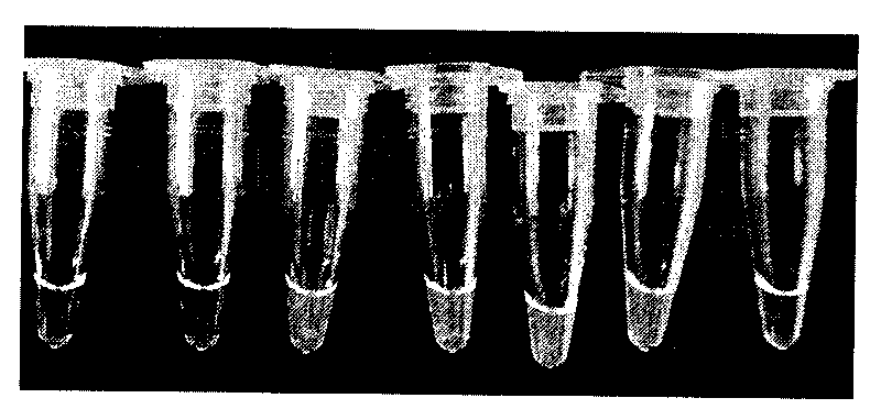
[0057] (4) After the amplification reaction, 15 μL of the system solution was taken, and 2 μL of 1000×SYBRGreen intercalator was directly added to the amplification tube, shaken and mixed, and the results were observed with the naked eye. Tubes without amplification reactions will be orange-yellow, and t...

Embodiment 3

[0059] (1) Reagent: Bst DNA polymerase large fragment produced by BioLabs (NEW ENGLAND) and 10 times Buffer solution; specific primer mixture; 4M betaine solution; 0.2M MgSO 4 solution;

[0060] (2) Amplification reaction system: the total volume of the amplification reaction is 25 μL, and its various components are: 10×Buffer 2.5 μL, 4M betaine 6.25 μL, 0.2M MgSO 4 , 0.25 μL of mixed primers, 3.5 μL of 10 μM dNTPs, 2 μL of 8000 U / L Bst DNA polymerase large fragment, 5 μL of template DNA, make up to 25 μL with sterilized deionized water, mix well and centrifuge;

[0061] (3) Amplification reaction procedure: proceed at 63°C for 45 minutes, and keep at 80°C for 2 minutes, store at 4°C;

[0062] (4) After the amplification reaction was completed, 25 μL of the system solution was taken and analyzed by 2% agarose gel electrophoresis, and the results were observed under ultraviolet light. Tubes without amplification reactions have no obvious bands, and tubes with amplification re...


PUM


Abstract

The invention discloses a method for rapidly detecting cow and sheep derived components. The principle is that a cow-derived forward outer primer COW-F3, a cow-derived backward outer primer COW-B3, a cow-derived forward inner primer COW-FIP, a cow-derived backward inner primer COW-FIP, a sheep-derived forward outer primer SHEEP-F3, a sheep-derived backward outer primer SHEEP-B3, a sheep-derived forward inner primer SHEEP-FIP, a sheep-derived backward inner primer SHEEP-BIP and DNA polymerase with the chain displacement activity are used for amplifying nucleic acid at 63-65 DEG C with the amplification efficiency up to 109-1,010 copies in a short time. The identifying method is to directly observe the turbidity of precipitation in a reaction tube by using naked eye or judge that amplification is carried out or not by the color change of added SYBR Green. The detection method has the characteristics of high specificity, high efficiency, rapidness, simple operation, easy detection and the like.

Description

technical field [0001] The invention belongs to the technical field of molecular biology, and relates to a rapid detection method for animal-derived components, more specifically, a method for rapidly detecting cattle and sheep-derived components, by visually observing the turbidity of the reaction tube or observing the color after adding 1000×SYBRGreen Change or observe the agarose gel electrophoresis to judge the amplification. Background technique [0002] Mad Cow disease (Mad COW disease) first appeared in Ashford Farm, England in 1985 as a "suspicious case". In November 1986, researchers conducted a histopathological examination on the diseased cow and found many spongy holes in the brain tissue, which was named Bovine spongiform encephalopathy (Bovine spongiform encephalopathy, BSE), and confirmed the disease as a new disease of cattle, first reported by Wells et al. in 1987. The prevalence of mad cow disease has caused serious impact and loss on the world's cattle in...

Claims


Application Information

Patent Timeline
Patent Type & Authority Applications(China)
IPC IPC(8): C12Q1/68C12N15/11
Inventor 朱珠王永兰青阔赵新程奕
Owner CENT LAB TIANJIN ACADEMY OF AGRI SCI
Why Eureka
Try Eureka