Method and device for three-dimensional ultrasonic real-time imaging and imaging system

An ultrasound imaging and equation technology, applied in image data processing, 2D image generation, instruments, etc., can solve the problems of increasing processing time and space overhead, the influence of light synthesis results, and the decrease of imaging speed, etc., to achieve real-time and three-dimensional ultrasound Imaging, high-quality three-dimensional ultrasound imaging, and the effect of improving imaging speed

Active Publication Date: 2010-07-07
SHENZHEN MINDRAY BIO MEDICAL ELECTRONICS CO LTD
View PDF0 Cites 21 Cited by
  • Summary
  • Abstract
  • Description
  • Claims
  • Application Information

AI Technical Summary

Problems solved by technology

The depth of error for each ray may vary, although the depth is always smaller than the ray step size, it still has some influence on the composite result of the ray
With the spatial distribution of light, the error depth shows a certain periodicity, resulting in ring, mesh, jagged and other specific shapes of texture in the imaging result, that is, the phenomenon of aliasing (Aliasing) occurs, which seriously affects the imaging quality
By setting a smaller ray step size, the aliasing phenomenon caused by the error depth can be alleviated, but it ...

Method used


Image

  • Method and device for three-dimensional ultrasonic real-time imaging and imaging system
  • Method and device for three-dimensional ultrasonic real-time imaging and imaging system
  • Method and device for three-dimensional ultrasonic real-time imaging and imaging system

Examples


Embodiment Construction

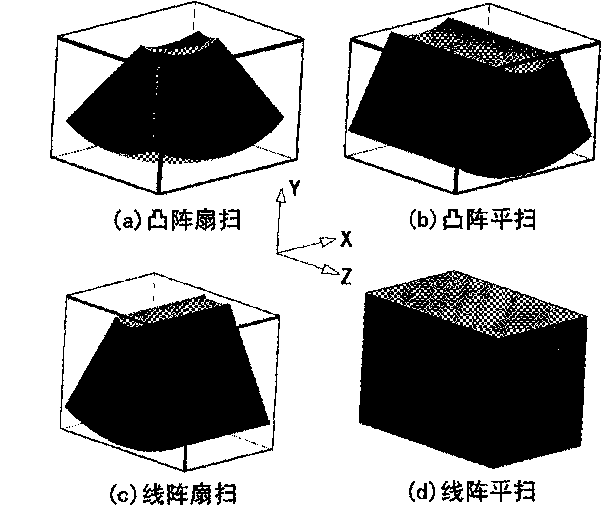
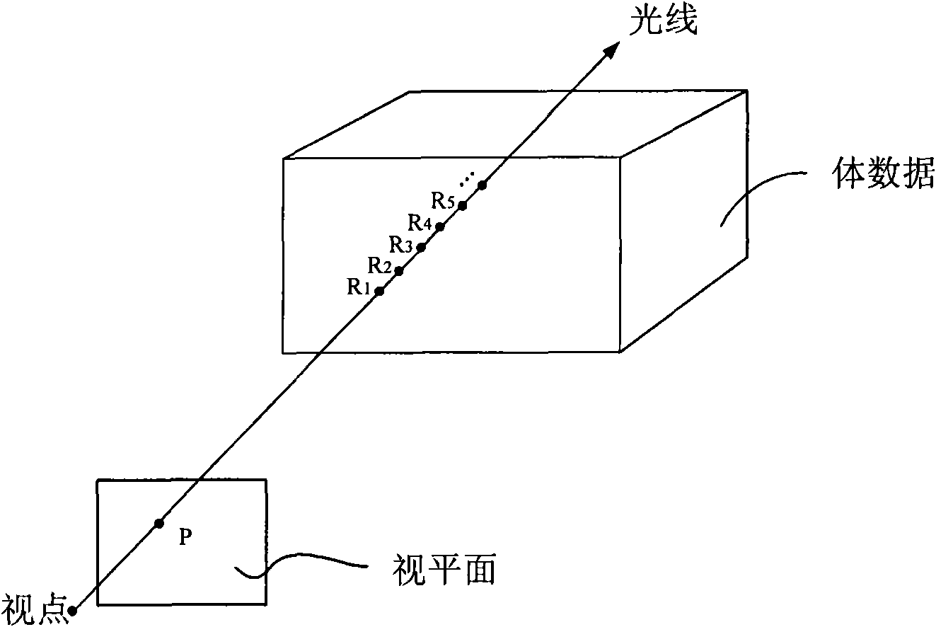
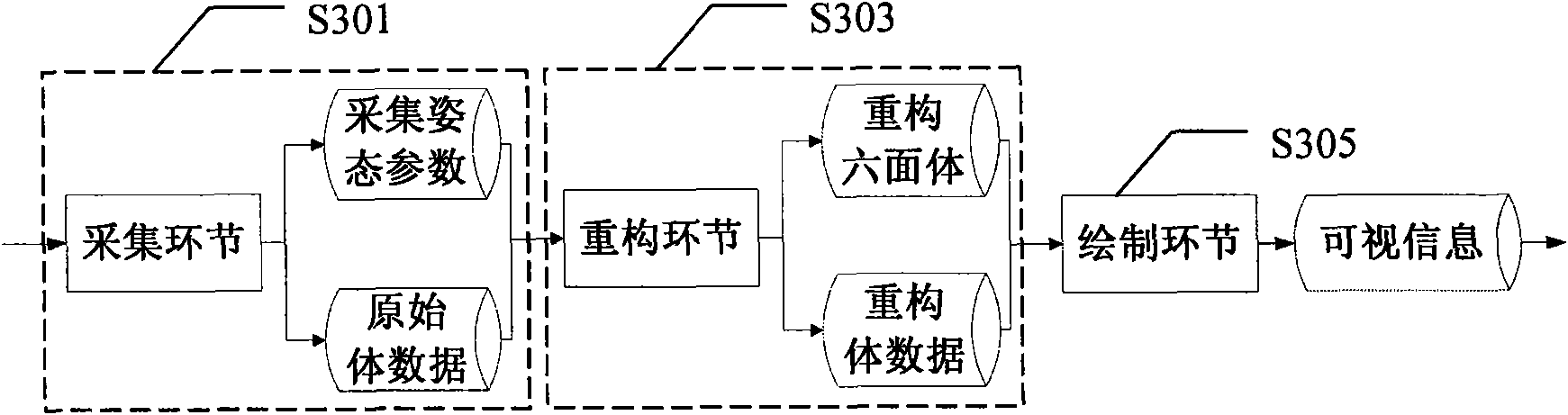
[0038] The three-dimensional ultrasound imaging process mainly includes three links: acquisition, reconstruction and rendering. The so-called acquisition (Acquisition) is the process of obtaining three-dimensional ultrasound volume data (Volume). At present, there are two main methods: the first is to use free-hand (Freehand) scanning, that is, hold a common probe and drag it along the thickness direction of the probe at a uniform speed. Moving or fanning at a constant speed to obtain a series of two-dimensional ultrasound images whose spatial position relationship can be estimated, and then obtain three-dimensional volume data offline; the other requires a special volumetric probe for scanning. Acquire a series of two-dimensional ultrasound images whose spatial position relationship can be determined, so as to obtain real-time three-dimensional volume data. The collected volume data consists of sequentially arranged voxels, and each voxel represents a point at a specific posi...


PUM


Abstract

The invention discloses a method and a device for three-dimensional ultrasonic real-time imaging and an ultrasonic imaging system comprising the device. A reconstructed polyhedron of body data is expressed by an analytical method and an imaging method, so that light rays can be cut to the range of the reconstructed polyhedron quickly and accurately, and the real-time and high-quality three-dimensional ultrasonic imaging can be realized.

Description

technical field [0001] The invention relates to an imaging method and device, in particular to a three-dimensional ultrasonic real-time imaging method and device, and an ultrasonic imaging system including the device. Background technique [0002] Traditional medical imaging equipment only provides two-dimensional images of the inside of the human body. Doctors can only estimate the size and shape of the lesion from multiple two-dimensional images based on experience, so as to imagine the three-dimensional geometric relationship between the lesion and its surrounding tissues, which brings great benefits to the treatment. difficulty. The 3D visualization technology can reconstruct a 3D shape from a series of 2D images and display it on the terminal. Therefore, not only can the overall concept of the intuitive and image of the imaging object be obtained, but also a lot of important three-dimensional information can be preserved. In addition, because ultrasound imaging has ob...

Claims


Application Information

Patent Timeline
IPC IPC(8): G06T11/00A61B8/13
Inventor 田勇佘跃姚斌胡勤军
Owner SHENZHEN MINDRAY BIO MEDICAL ELECTRONICS CO LTD
Why Eureka
Try Eureka