Medical and industrial X-ray real-time stereoscopic imaging device

A stereoscopic imaging and X-ray technology, applied in X-ray equipment, material analysis using radiation, electrical components, etc., can solve the problems of increasing the radiation dose of patients and doctors, large range of movement of the mechanism, prolonging the operation time, etc., to achieve mechanical movement The effect of small area, long exposure time and high radiation dose

Inactive Publication Date: 2011-01-26
北京睿思厚德辐射信息科技有限公司 +1
View PDF7 Cites 15 Cited by
  • Summary
  • Abstract
  • Description
  • Claims
  • Application Information

AI Technical Summary

Problems solved by technology

However, it is very laborious and unintuitive for doctors or non-destructive testing personnel to synthesize three-dimensional information from the front view, top view and side view just like looking at engineering drawings.
Moreover, every time the image is updated, the X-ray imaging devi

Method used


Image

  • Medical and industrial X-ray real-time stereoscopic imaging device
  • Medical and industrial X-ray real-time stereoscopic imaging device
  • Medical and industrial X-ray real-time stereoscopic imaging device

Examples


Example Embodiment

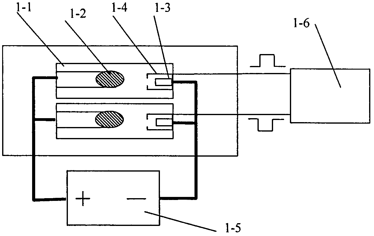
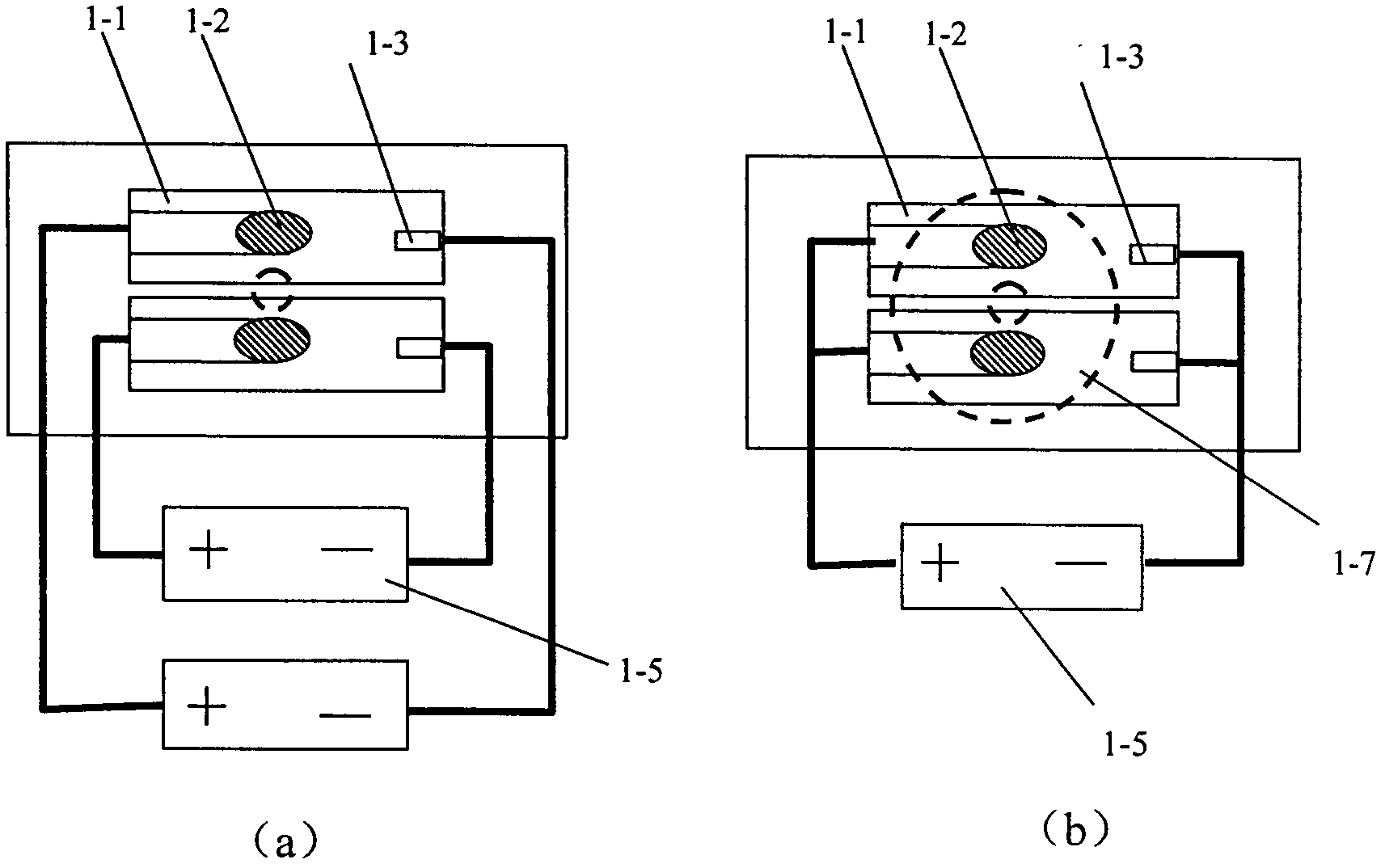
[0029] The medical and industrial X-ray real-time stereo imaging device proposed by the present invention has a structure such as figure 1 Shown, including:

[0030] The dual-view X-ray source 1 is used to generate X-rays 2 and 3 with two views, and the X-rays perform stereo imaging of the detection target;

[0031] The X-ray image detector 4 is used to detect the X-ray perspective image of the object to be measured and send the detected image to the computer. The X-ray image detector and the computer are connected by a signal line;

[0032] The X-ray machine moving platform is used to move the dual-view X-ray source and X-ray image detector around the detection target. The dual-view X-ray source is fixed on one side of the X-ray machine platform. The X-ray image detector is fixed on the other side of the X-ray machine platform opposite to the detection target;

[0033] The computer 5 is used for driving perspective transformation and synchronously acquiring perspective images of two ...


PUM


Abstract

The invention relates to a medical and industrial X-ray real-time stereoscopic imaging device, and belongs to the technical field of ray imaging equipment. The device comprises an X-ray source, an X-ray image sensor, a computer, a stereoscopic display and an X-ray machine stand, wherein the X-ray source has double viewing angles and can rapidly alternately emits X rays of double viewing angles; the X-ray image sensor is used for receiving X-ray images and realizing digitization; the computer can synchronously acquire images of left and right eyes and perform stereoscopic processing; the stereoscopic display is used for observing stereoscopic images; and the X-ray machine stand makes the X-ray source and the X-ray image sensor move or rotate in certain distance. The imaging device can display a three-dimensional perspective image of a human body or an object in real time during operation and perform three-dimensional positioning on organs and surgical instruments through a three-dimensional coordinate and a three-dimensional mouse, and also can display an X-ray three-dimensional image and the three-dimensional positioning of inner components of a tested object in real time in nondestructive testing or explosion-proof security. The imaging device has the advantages of high real-time, low cost and low exposure dose.

Description

technical field [0001] The invention relates to a medical and industrial X-ray real-time three-dimensional imaging device, which belongs to the technical field of medical and non-destructive detection radiographic imaging. Background technique [0002] X-ray imaging equipment has been widely used in medical diagnosis, minimally invasive surgery and industrial non-destructive testing. At present, X-ray imaging equipment produced at home and abroad, except for CT, is mostly a planar imaging system, which acquires two-dimensional plane perspective images, and cannot recognize the depth structure of a human body or an object. The positional relationship between surgical instruments and human organs involved in the human body is of great importance to precise treatment; industrial non-destructive testing also requires the detection of deep structure information. In principle, CT technology can be used for accurate three-dimensional imaging and positioning, but CT equipment is bu...

Claims


Application Information

Patent Timeline
IPC IPC(8): A61B6/03G01N23/04H05G1/08
Inventor 王经瑾宋征
Owner 北京睿思厚德辐射信息科技有限公司
Why Eureka
Try Eureka