Method for separating cells from different tissues

A tissue and source technology, applied in the field of separating cells from different tissue sources, can solve problems such as difficult to grasp the time of digestion, inability to observe tissue digestion, slow cell growth, etc., to achieve high practical application value, easy to quickly grasp, and uniform stress on cells Effect

Inactive Publication Date: 2011-06-15
FIELD OPERATION BLOOD TRANSFUSION INST OF PLA SCI ACAD OF MILITARY
View PDF1 Cites 7 Cited by
  • Summary
  • Abstract
  • Description
  • Claims
  • Application Information

AI Technical Summary

Problems solved by technology

[0003] Isolation of primary cells through mechanical digestion of tissue requires only simple surgical instruments, and the method is simple, easy to operate, and easy to master. However, during the process of mechanically separating tissue, the cells need to withstand a large mechanical force. Therefore, Isolated cells tend to grow slowly and take longer to recover
In addition, the mechanically trimmed tissue is not easy to digest completely during the digestion process, so that the number of cells obtained is extremely small
[0004] Isolation of tissue-derived cells by perfusion, although the tissue can be fully digested, but this method requires more surgical instruments, the method is difficult to master, and requires long-term practice
In addition, because collagenase stays in the tissue, it is impossible to observe the digestion of the tissue, and it is difficult to grasp the time of digestion
However, cells isolated using this method are in better condition

Method used


Image

  • Method for separating cells from different tissues
  • Method for separating cells from different tissues
  • Method for separating cells from different tissues

Examples


Embodiment 1

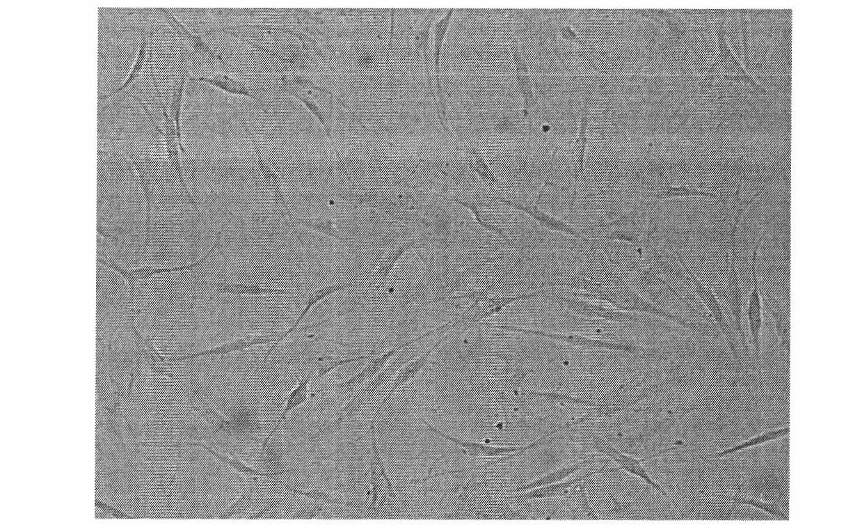
[0020] Embodiment 1, separation of adipose tissue primary cells

[0021] Taking adipose tissue (the adipose tissue is derived from the tissue taken out in the liposuction operation) as an example, the primary cells of the adipose tissue are separated by the method of the present invention, and the specific method comprises the following steps:

[0022] 1) Add 50mL of liposuction tissue into a sterile 50mL centrifuge tube, centrifuge at 1300rpm for 2 minutes, and suck out the lower liposuction anesthetic after centrifugation;

[0023] 2) Wash the adipose tissue with an equal volume of phosphate-buffered saline (PBS, pH 7.3, Gibcol), centrifuge at 1300 rpm for 2 minutes, suck out the lower layer of washing solution and discard it;

[0024] 3) Add Collagenase IV collagenase (sigma company), and adjust the collagenase concentration to 0.075% (V / V, 0.05%-0.1%), then add 2% (V / V, 1%-3% Yes) BSA 1.5mL (1-2mL is acceptable) and 0.2% (V / V, 0.1%-0.3% is acceptable) penicillin and strep...

Embodiment 2

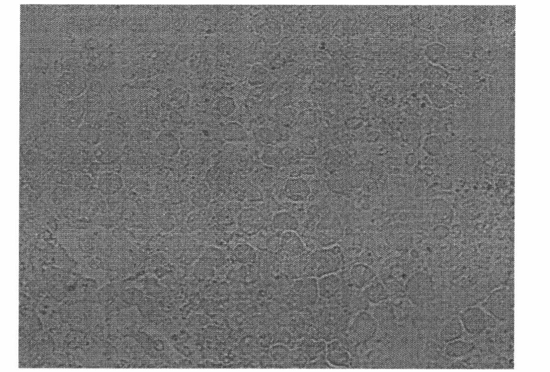
[0034] Embodiment 2, separation of liver tissue primary cells

[0035] Liver tissue acquisition: Take out a SD rat 2-3 days after birth (purchased from the Animal Experiment Center of the Academy of Military Medical Sciences of the Chinese People's Liberation Army) from the rat basin with a pair of large tweezers, and place it in 0.1% bromogeramine (purchased from Dezhou Lekang Disinfection Products Co., Ltd.) solution for 20-30s, take it out and place it on a foam board, fix the mouse with a needle (position it in a straight line), use another pair of large tweezers to take an iodine cotton ball to wipe the skin, and then rub it with alcohol. For cotton ball deiodination, take a pair of small scissors and tweezers to open the skin, fully tear it apart, then disinfect it with alcohol cotton balls, and open the abdominal cavity to take the liver.

[0036] Then put the removed liver in a round dish with DMEM medium prepared in the following step 1), remove the hemagglutination a...

Embodiment 3

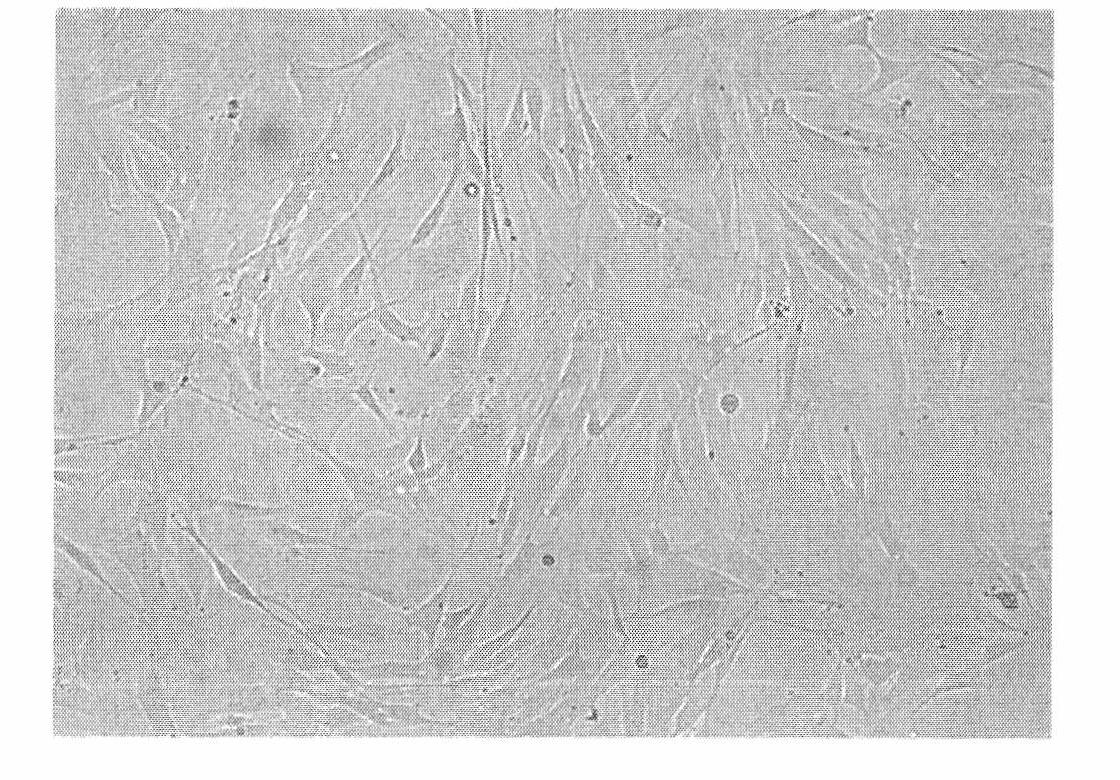
[0049] Example 3. Isolation of primary stromal cells derived from fetal liver tissue

[0050] Taking fetal liver tissue (fetal liver tissue is derived from aborted fetus) as an example, the primary cells of adipose tissue are separated by the method of the present invention. The specific method comprises the following steps:

[0051] 1) Insert two 5mL syringes into DMEM medium (purchased from Gibcol) and Collagenase IV collagenase (purchased from Gibcol) respectively, and add 5mL of DMEM culture solution to the two round dishes respectively.

[0052] 2) Add Collagenase IV Collagenase (Sigma Company) to the organized plate, and adjust the collagenase concentration to 0.1% (V / V, 0.05%-0.1%), then add 3% (V / V , 1%-3% can be) BSA 2mL (1-2mL can be) and 0.3% (V / V, 0.1%-0.3% can be) penicillin and streptomycin (purchased from Gibcol).

[0053] 3) Put the fetal liver tissue mixed with Dispase enzyme (please provide another available collagenase) in a triangular beaker (Indal Keyi Ma...


PUM


Abstract

The invention discloses a method for separating cells from different tissues. The method comprises the following steps of: placing tissues mixed with collagenase into a container in which a magnetic stirring rod is arranged; placing the container on a magnetic stirrer; switching on a power supply of the magnetic stirrer; adjusting the temperature to be between 35 and 37 DEG C at the rotation speed of 60 to 200rpm; and digesting the tissues at the temperature of between 35 and 37 DEG C and the rotation speed of 60 to 200rpm for 10 to 40 minutes to obtain tissue primary culture cells. The method has the advantages that: 1) the required apparatuses are simple, and the method is simple and is easy to operate and quick in grasping; 2) the state of the digested tissues can be observed in the whole process, the digestion degree of the collagenase is easy to grasp, and the separated cells have a better state; and 3) the tissues are fully digested through magnetic stirring better, the cells are uniformly stressed and are difficult to damage because the generated shearing force is relatively weak, and the cells obtained through separation have the better state.

Description

technical field [0001] The invention relates to a method for isolating specific cells, in particular to a method for isolating cells from different tissue sources. Background technique [0002] At present, there are mainly mechanical methods and perfusion methods for isolating primary cells from tissues, but both methods have obvious advantages and disadvantages. [0003] Isolation of primary cells through mechanical digestion of tissue requires only simple surgical instruments, and the method is simple, easy to operate, and easy to master. However, during the process of mechanically separating tissue, the cells need to withstand a large mechanical force. Therefore, Isolated cells tend to grow slowly and take longer to recover. In addition, mechanically trimmed tissues are difficult to digest completely during the digestion process, resulting in very few cells. [0004] Although the tissue-derived cells can be separated by perfusion, the tissue can be fully digested, but t...

Claims


Application Information

Patent Timeline
Patent Type & Authority Applications(China)
IPC IPC(8): C12N5/077C12N5/071
Inventor 裴雪涛滕越王韫芳南雪白慈贤师伟
Owner FIELD OPERATION BLOOD TRANSFUSION INST OF PLA SCI ACAD OF MILITARY
Why Eureka
Try Eureka