Mouse liver tumor cell line for highly expressing CD133 and preparation method thereof
A cell line and liver tumor technology, applied in biochemical equipment and methods, embryonic cells, animal cells, etc., can solve the problem of scarcity of tumor stem cells
- Summary
- Abstract
- Description
- Claims
- Application Information
AI Technical Summary
Problems solved by technology
Method used
Image
Examples
Embodiment 1
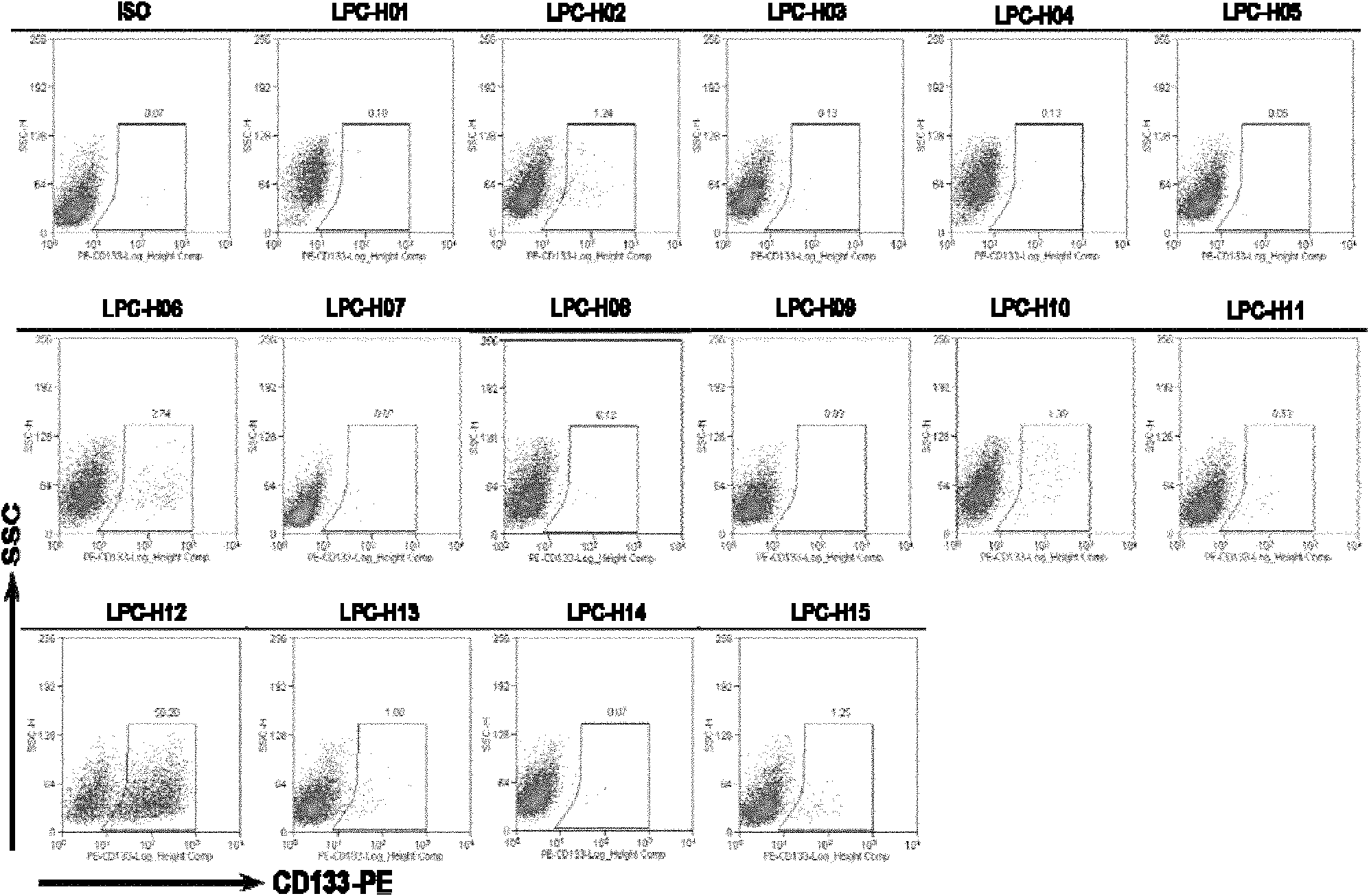
[0036] Establishment of mouse liver tumor cell line LPC-H12 with high expression of CD133:
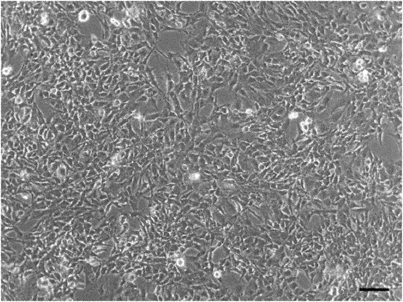
[0037] a) Isolation of p53- / - mouse embryonic liver cells (LPC cells): take p53- / - mouse embryos at E13.5d, rinse them in PBS for 2-3 times; use sterile ophthalmic scissors and tweezers to cut them completely liver. After cutting it into pieces, add liver digestion solution to digest at 37°C for 30 minutes to form a single cell suspension. Centrifuge to remove the supernatant, wash 2-3 times with PBS, and incubate the cells with rat anti-mouse E-cadherin monoclonal antibody at 4°C for 30min (the amount of antibody used is 4ug E-cadherin / 1×10 7 cells). Wash 2-3 times with PBS, centrifuge to remove the supernatant, and mix the cells with goat anti-rat magnetic beads (20ul / l×10 8 cells) were incubated at 4°C for 15 min. Finally, the E-cadherin+ embryonic liver cells were isolated by magnetic bead sorting system (MACS). The isolated cells were inoculated on a cell culture dish pre-coa...
Embodiment 2
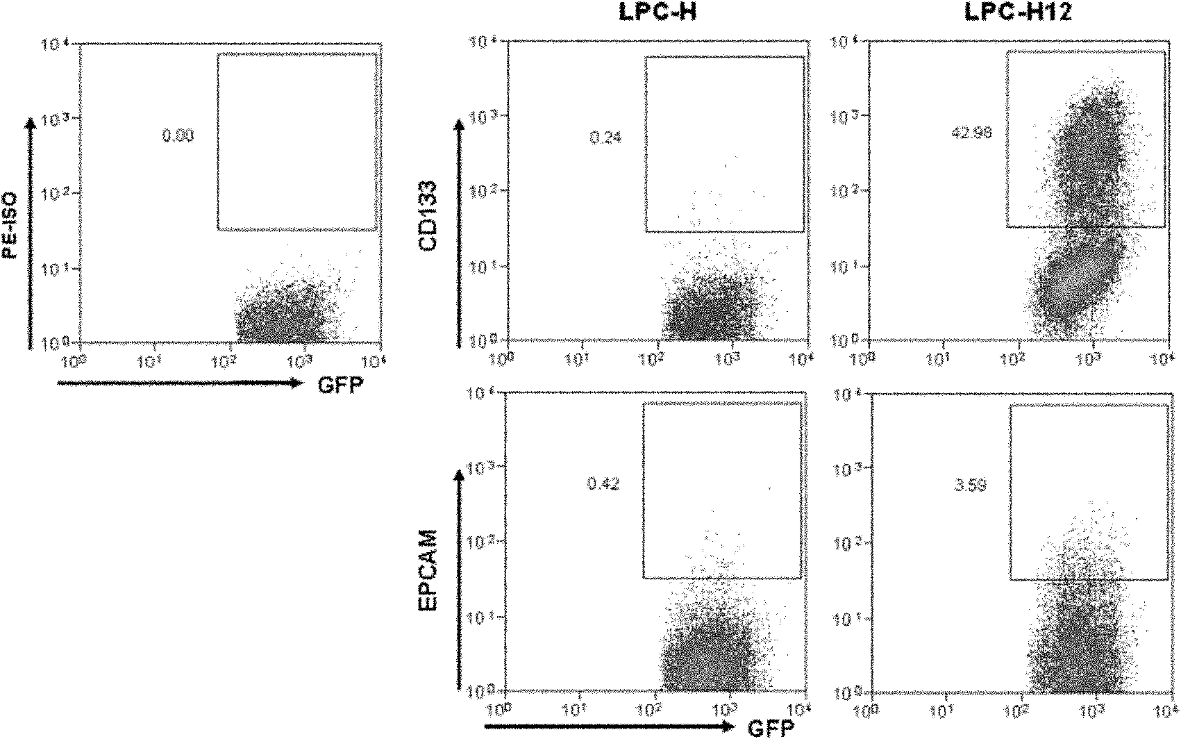
[0042] LPC-H12 cell line highly expresses stem cell markers CD133 and EpCAM:
[0043] a) The conventional in vitro culture conditions of LPC-H12 cell line are: DMEM / F12 culture medium prepared by 10% FBS and 50 mM 2-mercaptoethanol, 37°C, 5% carbon dioxide / 95% air, saturated humidity.
[0044] b) LPC-H12 cells grown to the logarithmic growth phase were digested with 0.25% trypsin+0.1% EDTA, washed twice with PBS, and 1×10 5 ~1×10 7 Put the cells into flow tubes, centrifuge, remove the supernatant, add 100 μl of 10% goat serum to suspend the cells, and block at 4° C. for 10 min.
[0045] c) Add CD133-PE or EpCAM-PE fluorescent antibody 2.5ul (0.5mg / ml) in step b), and incubate at 4°C for 30min.
[0046] d) Add FACS buffer, mix gently, centrifuge at 300g for 5min, and discard the supernatant.
[0047] e) Add 300-500 ul of FACS buffer solution, and perform detection with a BD flow analyzer. Such as image 3 Shown: LPC-H12 has higher expression levels of stem cell markers CD1...
Embodiment 3
[0049] Expression of liver stem cell-related genes in LPC-H12 cell line:
[0050] a) The LPC-H12 cells that were routinely cultured in vitro and grown to the logarithmic growth phase were digested with 0.25% trypsin + 0.1% EDTA, and then the cells were collected. The cells were washed twice with PBS, and the total RNA extraction kit from TaKara was used for the cell pellet. Extraction of total RNA was performed.
[0051] b) Take 2ug of total RNA and reverse it into cDNA using the reverse transcription kit from TaKara Company.
[0052] c) PCR detection of expression of genes related to proliferation and self-renewal of hepatic stem cells. The amplified target gene and its primer sequences are shown in Table 1, and the primers were synthesized by Handsome Biotechnology Co., Ltd. The PCR reaction conditions were pre-denaturation at 95°C for 5 min, and cycled 35 times. The cycle conditions were: denaturation at 95°C for 130 s, annealing for 30 s, the annealing temperature is sho...
PUM
 Abstract
Description
Claims
Application Information
 - R&D
- Intellectual Property
- Life Sciences
- Materials
- Tech Scout
- Unparalleled Data Quality
- Higher Quality Content
- 60% Fewer Hallucinations
Browse by: Latest US Patents, China's latest patents, Technical Efficacy Thesaurus, Application Domain, Technology Topic, Popular Technical Reports.
© 2025 PatSnap. All rights reserved.Legal|Privacy policy|Modern Slavery Act Transparency Statement|Sitemap|About US| Contact US: help@patsnap.com


