Caspase-3 proenzyme protein immunohistochemical diagnosis kit and application

A technology of caspase and diagnostic kit, which is applied in the direction of measuring devices, instruments, scientific instruments, etc., can solve the problem of high non-specificity, cumbersome operation procedures, and the inability to quickly and accurately judge the expression of Procaspase-3 antigen in tumor tissue of patients to achieve the effect of high specificity and simple operation procedures

Inactive Publication Date: 2011-09-14
SHENYANG PHARMA UNIVERSITY
View PDF3 Cites 8 Cited by
  • Summary
  • Abstract
  • Description
  • Claims
  • Application Information

AI Technical Summary

Problems solved by technology

[0005] Since there are no Procaspase-3 protein immunohistochemical diagnostic kits at home and abroad, the existing methods for detecting Procaspase-3 protein in tissues have cumbersome procedures and high non-specificity, and cannot quickly and accurately determine the Procaspase-3 antigen in tumor tissues of patients. expression of

Method used


Image

  • Caspase-3 proenzyme protein immunohistochemical diagnosis kit and application
  • Caspase-3 proenzyme protein immunohistochemical diagnosis kit and application

Examples


experiment Embodiment 1

[0030] Experimental Example 1 Composition of Procaspase-3 Protein Immunohistochemical Diagnostic Kit

[0031] Table 1 Product composition and main components of 40 servings / box

[0032]

experiment Embodiment 2

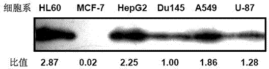
[0033] Experimental example 2 Selection of tumor cells with different expression levels of Procaspase-3 antigen

[0034] Tumor cell selection was performed using the following steps:

[0035] 1) Alternative tumor cells include:

[0036] Human liver cancer cell line HepG2, human lung cancer cell line A549, human breast cancer cell line MCF-7, human glioma cell line U87, human prostate cancer cell line DU145 and human leukemia cell line HL-60 (purchased from ATCC). After culturing in vitro to the logarithmic growth phase, various cells were collected for cell technology.

[0037] 2) Protein extraction

[0038] Grow to 10 7Cells were centrifuged at 1000 rpm for 5 min to collect various cells, washed once with PBS, centrifuged again, and the supernatant was discarded. Add 100 μL of pre-cooled cell lysate to each cell, lyse the sample at 4°C for 30 min, centrifuge at 12,000 rpm for 15 min, and take the supernatant for use. Store at -80°C.

[0039] 3) Preparation of working so...

experiment Embodiment 3

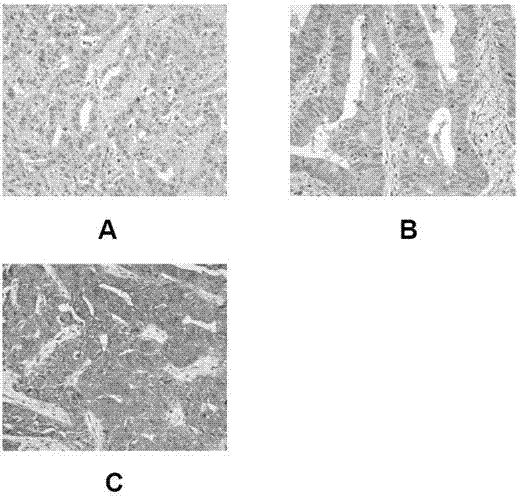
[0069] Experimental example 3 Preparation of tumor tissues with different expression levels of Procaspase-3

[0070] 1) Select HepG2, DU145, and MCF-7 tumor cells with high, moderate, and no expression of Procaspase-3 obtained in Example 1, and passage them in RPMI-1640 complete medium containing 10% fetal bovine serum amplify;

[0071] 2) For the three types of cells in the logarithmic growth phase, wash with PBS, centrifuge, discard the supernatant, resuspend the cell pellet in PBS, count, and adjust the cell density to 2x10 7 / mL;

[0072] 3) The tumor cell suspension was inoculated into the left and right armpits of BALB / C nude mice, and the tumor growth in the inoculated area was regularly observed;

[0073] 4) When the tumor grows to 300mm 3 Left and right size (tumor volume calculation formula is X 2 y / 2, where X is the long diameter of the tumor and y is the short diameter of the tumor), the animals were sacrificed by cervical dislocation, and the tumor tissues we...


PUM


Abstract

The invention discloses a procaspase-3 immunohistochemical diagnosis kit and a detection method thereof. The kit comprises an immunohistochemical staining agent, which comprises an initial antibody, a second antibody, enzyme, a substrate, a color-developing agent, a blocking agent and buffer solution, wherein the initial antibody is connected with tissue antigen; the second antibody is connected with the initial antibody; and the blocking agent is positioned at a non-specific binding site. The kit can measure expression level of the procaspase-3 antigen and cell position condition in the testee tissue accurately, provides guidance for clinical application of medicaments using the procaspase-3 protein as a target spot, and provides reference for clinical diagnosis of malignant tumor, hepatic fibrosis, renal injury and neurodegenerative lesion. The invention also discloses a control picture for detecting expression level of the procaspase-3 protein. The control picture is provided with a procaspase-3 high-expression control region, a procaspase-3 medium-expression control region, a procaspase-3 non-expression (low-expression) control region and a procaspase-3 detection region for placing the tissue to be detected. The control picture provides an accurate evaluation criterion for evaluation of the expression level of the procaspase-3.

Description

technical field [0001] The invention relates to an SP method immunohistochemical kit and its application, which mainly consist of a procaspase-3 zymogen (Procaspase-3) monoclonal antibody, a biotinylated secondary antibody, a chromogenic solution and a comparison photo. Background technique [0002] Procaspase-3 is the precursor zymogen of Caspase-3, which plays a crucial role in the transmission of apoptosis signals. Procaspase-3 not only receives cascade stimulation of exogenous apoptotic pathway, but also is activated by endogenous apoptotic pathway. Procaspase-3 is the most downstream molecule of the entire apoptotic pathway, so once its abnormal expression or function changes, the entire apoptotic signaling pathway will be blocked or activated. [0003] At present, more and more literatures have reported that apoptosis is closely related to many diseases. It has been confirmed that one of the causes of neurodegenerative diseases is the apoptosis of neurons in certain ...

Claims


Application Information

Patent Timeline
Patent Type & Authority Applications(China)
IPC IPC(8): G01N33/577
Inventor 吴春福杨静玉王立辉宫平
Owner SHENYANG PHARMA UNIVERSITY
Why Eureka
Try Eureka