DICOM (Digital Imaging and Communications in Medicine) medical image encrypting and decrypting method based on edge detection

An edge detection and medical image technology, applied in the field of image processing, can solve the problems of slow calculation speed, good confidentiality, loss of image information, etc., and achieve the effect of reducing the amount of encrypted data, reducing the amount of calculation, and improving efficiency

Active Publication Date: 2011-09-14
CHONGQING UNIV
View PDF3 Cites 10 Cited by
  • Summary
  • Abstract
  • Description
  • Claims
  • Application Information

AI Technical Summary

Problems solved by technology

[0004] The disadvantages of the existing technology are: the medical image has a large amount of data, the pixel structure is strong, and distortion is not allowed, but the current encryption method is either slow in calculation speed and confidential Insufficient performance, or the image information will be lost, resulting in misjudgment of the disease, and cannot meet the requirements of fast computing speed, good confidentiality, and no distortion at the same time

Method used


Image

  • DICOM (Digital Imaging and Communications in Medicine) medical image encrypting and decrypting method based on edge detection
  • DICOM (Digital Imaging and Communications in Medicine) medical image encrypting and decrypting method based on edge detection
  • DICOM (Digital Imaging and Communications in Medicine) medical image encrypting and decrypting method based on edge detection

Examples


Embodiment 1

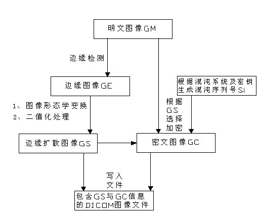
[0046] The plaintext image GM of the present embodiment is a skull image with a size of 256×256 from the collection of CASIMAGE DICOM sample graphics files, such as image 3 shown.

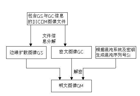
[0047] like figure 1 Shown: a kind of DICOM medical image encryption and decryption method based on edge detection comprises encryption process and decryption process, and described encryption process is carried out in the following steps:

[0048] step one, yes image 3 Perform edge detection on the plaintext image GM shown, and obtain the edge image GE of the plaintext image GM, such as Figure 4 shown;

[0049] The edge detection method adopted is: Convolve the image with a 3*3 Laplace convolution kernel.

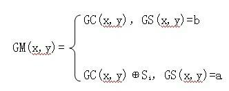
[0050] Step 2, using image morphological transformation to diffuse the edge in the edge image GE, and binarize the diffused image to obtain the edge diffusion image GS, the pixel value of the edge part of the edge diffusion image GS is a , and the rest of the pixel values ​​are b;

[0...

Embodiment 2

[0084] The plaintext image GM of the present embodiment is also a skull image with a size of 256×256 selected from the CASIMAGE DICOM sample graphics file collection, such as Figure 11 shown.

[0085] This embodiment is roughly the same as Embodiment 1, except that the adopted edge detection method is: use a square filter Sobel operator with a width of 3 to perform a first-order derivation on each of the horizontal and vertical axes; the adopted The image morphological transformation method is as follows: a 3*3 rectangular kernel with a reference point at the center performs an iterative expansion operation.

[0086] The plaintext image GM, edge image GE, edge diffusion image GS, and ciphertext image GC in the encryption process of this embodiment are as follows: Figure 11 , 12 , 13, and 14; the ciphertext image GC, edge diffusion image GS, and plaintext image GM in the decryption process are shown as Figure 15 , 16 , 17 shown. The key used in this embodiment is a=0.66...


PUM


Abstract

The invention discloses a DICOM (Digital Imaging and Communications in Medicine) medical image encrypting and decrypting method based on edge detection, comprising an encrypting process and a decrypting process. Based on a selective encryption idea, the method comprises the following steps of: firstly using an edge detection technology to calculate edge information of a plaintext image; amplifying the edge range through an image morphological exchange operation; and then encrypting and decrypting the pixel of the plaintext image corresponding to the edge through a chaos system. Compared with a traditional medical image encrypting method, by application of the method of the invention, key information in the medical images is excellently encrypted; the encrypting data amount is greatly reduced; the encrypting efficiency is improved; and the method has strong practicability.

Description

technical field [0001] The invention belongs to the field of image processing, in particular to a method for encrypting and decrypting DICOM medical images based on edge detection. Background technique [0002] DICOM (Digital Imaging Communication in Medicine) is an international standard widely followed in the construction of PACS (Picture Archiving and Communication Systems). The introduction of the DICOM standard greatly simplifies the realization of medical image information exchange, promotes the research and development of teleradiology systems, image archiving and communication systems, and makes it compatible with other medical application systems (Medical Information System HIS, Radiology Information Management System RIS) The integration of medical technology has become possible, bringing new opportunities for the digitalization of medicine. [0003] With the popularization of DICOM standard and the rise of telemedicine, how to encrypt medical images safely and ...

Claims


Application Information

Patent Timeline
Patent Type & Authority Applications(China)
IPC IPC(8): G06T1/00
Inventor 向涛罗小松屈晋宇余晨韵李传东
Owner CHONGQING UNIV
Why Eureka
Try Eureka