Separation method for cell cluster having tumorigenic potential in liver cancer tissue

A separation method and cell cluster technology, applied in the field of tumor tissue cells and primary culture of tumor tissue cells, can solve the problems of separation and identification of liver cancer stem cell subsets, difficult to obtain viable cell sorting and identification, etc., and achieve the effect of narrowing the search range.

Inactive Publication Date: 2013-05-29
ZHEJIANG UNIV
View PDF1 Cites 0 Cited by
  • Summary
  • Abstract
  • Description
  • Claims
  • Application Information

AI Technical Summary

Problems solved by technology

However, most clinical liver cancer samples are accompanied by severe liver fibrosis, and it is difficult to obtain vigor and sufficient number of cells to complete the sorting and necessary identification, and it is difficult to separate and identify cell subsets with liver cancer stem cell characteristics on this basis

Method used


Image

  • Separation method for cell cluster having tumorigenic potential in liver cancer tissue
  • Separation method for cell cluster having tumorigenic potential in liver cancer tissue
  • Separation method for cell cluster having tumorigenic potential in liver cancer tissue

Examples


Embodiment 1

[0089] Example 1: Sample basic information

[0090] All 36 specimens were taken from HBV-positive patients. The preoperative imaging diagnosis was all hepatocellular carcinoma patients with single tumor focus and clear border. The tumor diameter was 3.2cm-7.4cm (4.4±1.2cm), without vascular invasion and distant metastasis. There were varying degrees of fibrosis in the diseased livers.

Embodiment 2

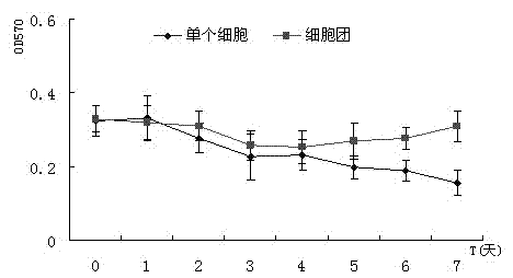
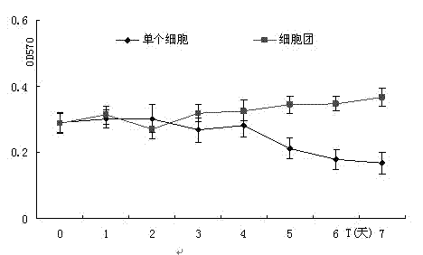
[0091] Example 2: Cell and Cell Aggregate Activity

[0092] After using the above separation method, the suspension obtained is a mixture of single cells and cell aggregates of different sizes. Trypan blue staining shows that the blue-stained cells are 6 (1.312±0.464*10 7 ), trypan blue staining showed that the viable cells in the obtained cell suspension accounted for 95.7±4.5%. Take part of the obtained small group of tissues and further digest them to count the number of cells and calculate the total number of cells. This part of the cells is included in the statistical results. The average number of cells per gram of sample processed by this method is 1.625±0.324*10 7 cell. The number of cells in the small tissue block is 9-29 (23.8±6.9), which can be attached to the surface of the petri dish coated with rat tail collagen after being diluted to an appropriate multiple with the medium. The results of trypan blue staining showed that the viable cells accounted for 96.0±3...

Embodiment 3

[0093] Example 3: Expression of different proteins in liver cancer tissues

[0094] The results of immunohistochemistry showed that the anti-human hepatocyte antibody was positive in the hepatocellular carcinoma cell membrane in all 36 cases of liver cancer samples; CK19 was strongly positive in the bile duct epithelial cells: in the liver cancer tissues, most of the liver cancer cells were CK18 positive, and in the The cytoplasm and membrane of some cells scattered around the central vein were strongly positive for anti-CK18; consistent with our previous research results [1] , CD133-positive cells scattered in cords or clusters in liver cancer cells.


PUM

PropertyMeasurementUnit
diameteraaaaaaaaaa

Abstract

The invention discloses a separation method for a cell cluster having a tumorigenic potential in liver cancer tissue. By using the method, the cell cluster having the capacities of growth and proliferation in vitro, colony forming and tolerance to anticancer drugs can be separated from a clinical sample quickly, easily and effectively, and the passage cells have certain stability and are the foundation of separating and identifying the source cells having high tumorigenicity. In the condition of studying liver cancer stem cell and micro-environment for liver cancer stem cell growth without enough stable cells that are hard to be obtained in primary tissue, the study of the composition of the cell clusters and the role of the cell clusters in maintaining the continuous increasing of tumor cells, resistance to drugs and tumorigenicity can help researchers to gradually narrow the search area, so as to finally lock the liver cancer stem cell that causing a liver caner and a recurrence after treatment, and combine the study of the micro-environment of the cell to explore a feasible scheme for clinic treatment of great reference value.

Description

technical field [0001] The invention belongs to the field of tumor tissue cells and primary culture of tumor tissue cells, and in particular relates to an in vitro method for obtaining liver cancer cell subpopulations with relatively stable properties, high growth and proliferation, tumorigenic ability and high drug resistance in liver cancer clinical specimens. Background technique [0002] At present, it is believed that tumor cells are heterogeneous, and a small number of cancer stem cells show stronger growth and proliferation, tumorigenicity and resistance to antitumor drugs, and are the source cells of tumor formation and maintenance and recurrence after treatment. Finding these cells and determining their cell biological characteristics will help to develop new diagnosis and treatment schemes, realize early diagnosis and targeted treatment schemes of malignant tumors, and reduce their incidence and lethality. [0003] In 1997, it was discovered that there is a subset ...

Claims


Application Information

Patent Timeline
Patent Type & Authority Patents(China)
IPC IPC(8): C12N5/09
Inventor 殷胜勇谢海洋周琳郑树森
Owner ZHEJIANG UNIV
Why Eureka
Try Eureka