Joint detection method for heart cerebrovascular disease-related protein marker and diagnostic kit thereof
A technology of cardiovascular and cerebrovascular diseases and protein markers, which is applied in the field of liquid phase chip combined with parallel detection method and its diagnostic kit, can solve the problem of poor reproducibility of solid phase biochip technology, limitation of detection throughput, insufficient sensitivity, etc. problems, to achieve high sensitivity, rapid detection, and good stability
- Summary
- Abstract
- Description
- Claims
- Application Information
AI Technical Summary
Problems solved by technology
Method used
Image
Examples
Embodiment 1
[0048] Example 1: Liquid-phase chip joint detection method for two kinds of protein markers related to cardiovascular and cerebrovascular diseases
[0049] The specific detection method includes the following steps:
[0050] Select 2 kinds of microspheres numbered 11 and 15
[0051] 1. Activation of desired microspheres
[0052] 1.1 Vortex the microsphere storage solution at full speed for at least 3 minutes to form a uniform microsphere suspension;
[0053] 1.2 Weigh 10 mg of EDC and S-NHS into two centrifuge tubes;
[0054] 1.3 Dissolve in deionized water to make the final concentration 50mg / ml;
[0055] 1.4 Take 1ml of the microsphere suspension and centrifuge at 10000g for 3min, carefully remove the supernatant;
[0056] 1.5 Add 80 μl of activation buffer to resuspend the microspheres;
[0057] 1.6 Add 10 μl of EDC solution (50 mg / ml) and 10 μl of S-NHS solution (50 mg / ml) respectively, mix well, and incubate at room temperature (15-25° C.) in the dark and shake for 2...
Embodiment 2
[0110] Example 2: Liquid-phase chip combined detection method for three kinds of protein markers related to cardiovascular and cerebrovascular diseases
[0111] The specific detection method includes the following steps:
[0112] Select 3 kinds of microspheres numbered 21, 33, and 37
[0113] 1-3 steps are the same as in Example 1.
[0114] 4. Configuration of Antigen Standards
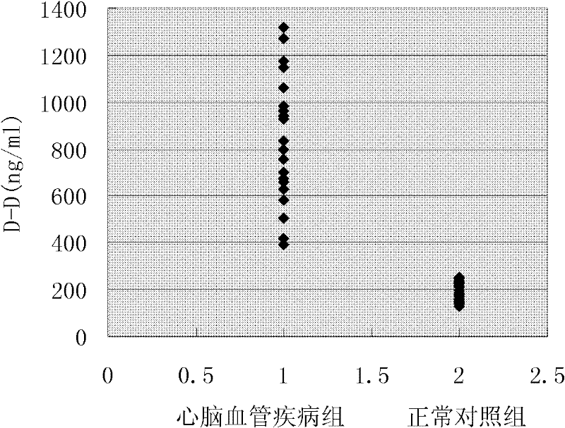
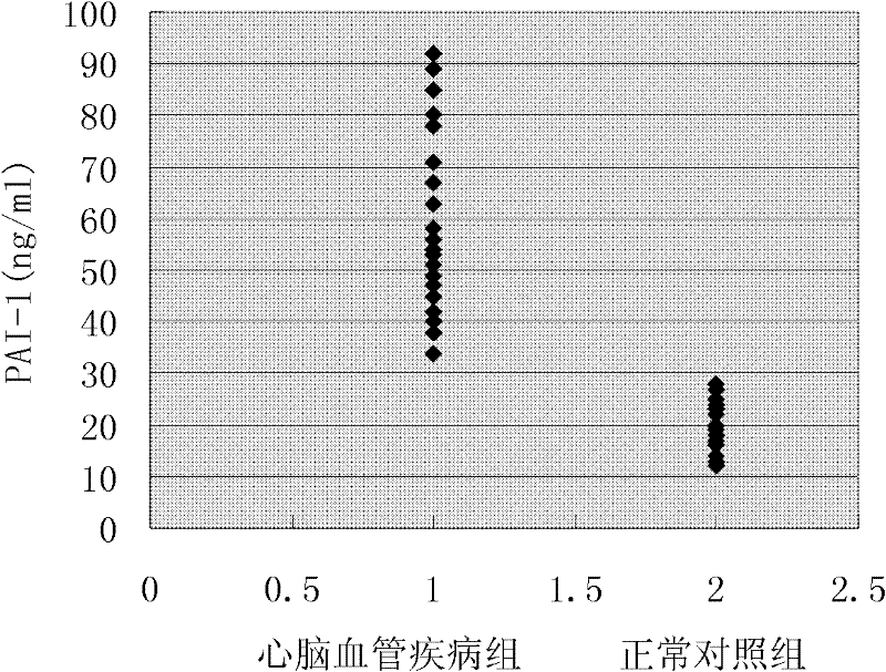
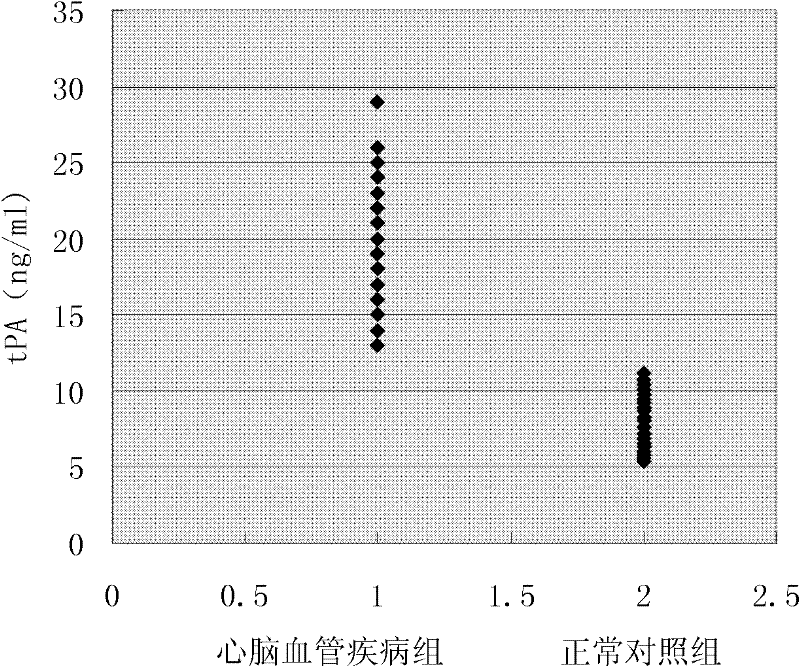
[0115] D-D was prepared according to the concentration of 31250, 6250, 1250, 250, 50, 10, 0ng / ml, PAI-1 and tPA were prepared according to the concentration of 312.5, 62.5, 12.5, 2.5, 0.5, 0.1, 0ng / ml, and the markers The mixtures are labeled as STD6, STD5, STD4, STD3, STD2, STD1, STD0, respectively.
[0116] 5. Preparation of the microsphere mixture (I mixture) coupled with the capture antibody.
[0117] Take the microspheres coated with the capture antibodies of three kinds of cardiovascular and cerebrovascular-related protein markers, as follows: D-D capture antibody microspheres 21, PAI-1 ca...
Embodiment 3
[0144] Example 3: Liquid chip combined detection method for 5 kinds of protein markers related to cardiovascular and cerebrovascular diseases
[0145] The specific detection method includes the following steps:
[0146] Select 5 kinds of microspheres numbered 11, 15, 21, 33, and 37
[0147] 1-3 steps are the same as in Example 1.
[0148] 4. Configuration of Antigen Standards
[0149] D-D is prepared according to the concentration of 31250, 6250, 1250, 250, 50, 10, 0ng / ml, PAI-1, LEP and tPA are prepared according to the concentration of 312.50, 62.50, 12.50, 2.50, 0.50, 0.10, 0ng / ml, ApoB was prepared at concentrations of 31.25, 6.25, 1.25, 0.25, 0.05, 0.01, and 0 mg / ml, and the marker mixtures were labeled as STD6, STD5, STD4, STD3, STD2, STD1, and STD0, respectively.
[0150] 5. Preparation of microsphere mixture (mixture I) coupled with capture antibody
[0151] Take the microspheres coated with capture antibodies of five kinds of protein markers related to cardiovas...
PUM
 Abstract
Description
Claims
Application Information
 - R&D
- Intellectual Property
- Life Sciences
- Materials
- Tech Scout
- Unparalleled Data Quality
- Higher Quality Content
- 60% Fewer Hallucinations
Browse by: Latest US Patents, China's latest patents, Technical Efficacy Thesaurus, Application Domain, Technology Topic, Popular Technical Reports.
© 2025 PatSnap. All rights reserved.Legal|Privacy policy|Modern Slavery Act Transparency Statement|Sitemap|About US| Contact US: help@patsnap.com


