Delivery starting forecasting device based on uterine electromyography and method for extracting characteristic parameter
An electromyography, uterine technology, applied in medical science, sensors, diagnostic recording/measurement, etc., can solve the problem of low degree of accuracy, reduce the incidence, reduce morbidity and mortality, reduce the risk of childbirth and newborns The effect of child defect rate
- Summary
- Abstract
- Description
- Claims
- Application Information
AI Technical Summary
Problems solved by technology
Method used
Image
Examples
Embodiment 1
[0040] Such as figure 1Shown is a labor initiation forecasting device based on uterine electromyography, including: uterine electromyography signal acquisition system, uterine electromyography digital signal processing system, graphic display device for processing results and sound forecasting device, uterine electromyography The signal acquisition system is connected with the digital signal processing system of the electromyogram of the uterus, and the digital signal processing system of the electromyography of the uterus is connected with the graphic display device and the sound forecasting device.
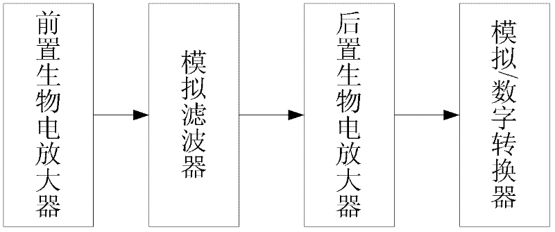
[0041] Such as image 3 As shown, the uterine electromyography signal acquisition system includes: electrodes for extracting pregnant women’s abdominal electrical signals containing uterine electromyography, and a pre-bioelectric amplifier for amplifying the extracted electrical signals without distortion. An analog filter for filtering out interference signals, a post bioelect...
Embodiment 2
[0053] Such as Figure 5 As shown, for the above-mentioned device for predicting the start of labor based on uterine electromyography, the characteristic parameters can also be extracted by a single acquisition method of pregnant women's uterine electromyography signal, which includes the following basic steps:
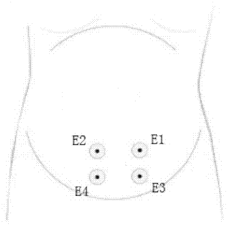
[0054] (1) In the first two weeks of the expected date of delivery, collect the EMG signal of the pregnant woman once. The electrodes are placed in the following positions: E1 is placed 3.5cm to the left of the navel and 1cm below the navel; E2 is placed 3.5cm to the right of the navel. 1cm below the navel; E3 was placed 3.5cm to the left of the navel and 8cm below the navel; E4 was placed 3.5cm to the right of the navel and 1cm below the navel; the reference electrode was placed on the hip. The uterine electromyography signal is collected for 30 minutes. Considering the comfort of pregnant women, the extraction time can also be shortened appropriately, and the differ...
PUM
 Abstract
Description
Claims
Application Information
 - R&D
- Intellectual Property
- Life Sciences
- Materials
- Tech Scout
- Unparalleled Data Quality
- Higher Quality Content
- 60% Fewer Hallucinations
Browse by: Latest US Patents, China's latest patents, Technical Efficacy Thesaurus, Application Domain, Technology Topic, Popular Technical Reports.
© 2025 PatSnap. All rights reserved.Legal|Privacy policy|Modern Slavery Act Transparency Statement|Sitemap|About US| Contact US: help@patsnap.com


