Superficial blood vessel display method and instrument

A display method and blood vessel technology, applied in sensors, medical science, diagnostic recording/measurement, etc., can solve problems such as accidental injury to patients, death, and bleeding of arterial and vascular patients, and achieve the effect of convenient operation

Active Publication Date: 2012-04-18
HEER MEDICAL TECH DEV CO LTD
View PDF3 Cites 21 Cited by
  • Summary
  • Abstract
  • Description
  • Claims
  • Application Information

AI Technical Summary

Problems solved by technology

[0006] There are following defects in the prior art: during some operations, under the premise that some patients are difficult to find veins, the medical staff accidentally injures the patient's arterial blood vessel, causing massive bleeding or even death of the patient

Method used


Image

  • Superficial blood vessel display method and instrument
  • Superficial blood vessel display method and instrument
  • Superficial blood vessel display method and instrument

Examples


Embodiment Construction

[0035] The technical solutions in the embodiments of the present invention will be clearly and completely described below in conjunction with the accompanying drawings in the embodiments of the present invention. It should be noted that the described embodiments are only part of the embodiments of the present invention, not all of them. Example. Based on the embodiments of the present invention, all other embodiments obtained by persons of ordinary skill in the art without making creative efforts belong to the protection scope of the present invention.

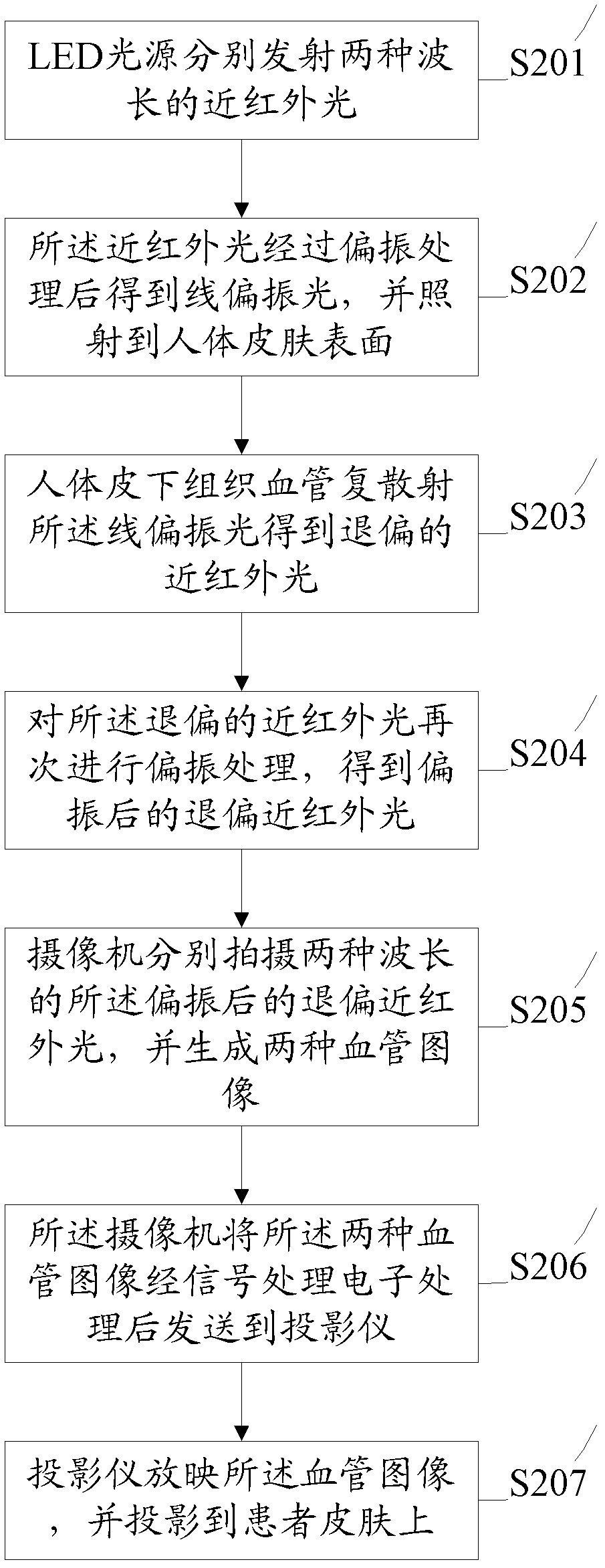
[0036] An embodiment of the present invention provides a method for displaying superficial blood vessels, such as figure 2 shown, including:

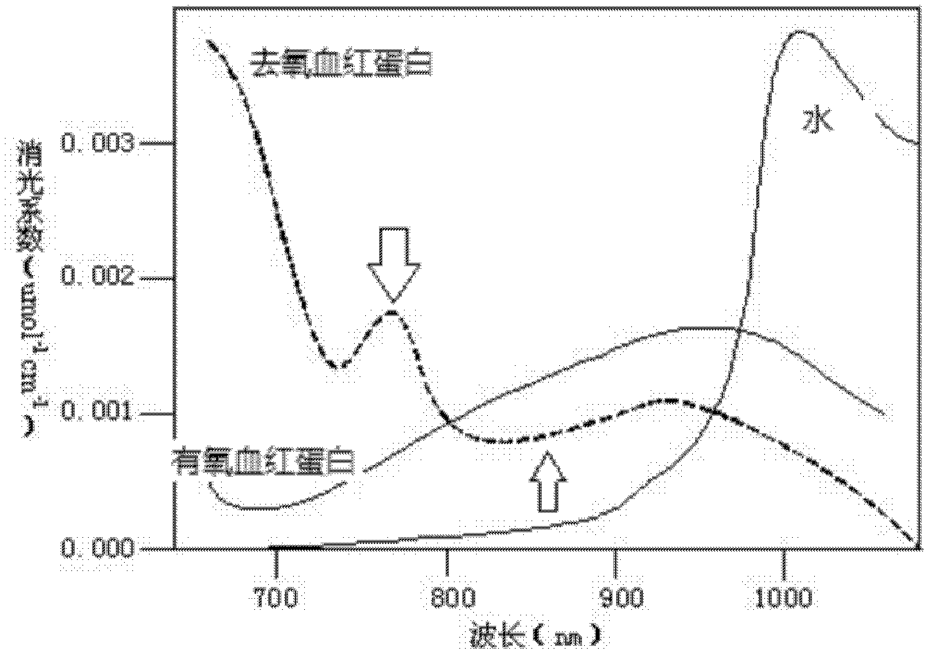
[0037] In step S201, the LED light source emits near-infrared light of two wavelengths respectively.

[0038] The LED light source emits near-infrared light of two specific wavelengths, and the LED light source is composed of two kinds of LED lamps with center wavelengths of 760nm an...


PUM


Abstract

The invention discloses a superficial blood vessel display method, which comprises the following steps that: an LED (light-emitting diode) light source respectively emits near infrared light with two wavelengths; the near infrared light is polarized to obtain linearly polarized light which irradiates human body skin surface; blood vessels of human body subcutaneous tissues carry out multi-scattering on the linearly polarized light to obtain depolarized near infrared light; the depolarized near infrared light is polarized again to obtain polarized depolarized near infrared light; a camera respectively shoots the polarized depolarized near infrared light with two wavelengths to generate two blood vessel images; the camera performs signal processing and electronic processing on the two blood vessel images and sends the processed images to a projector; and the projector shows the blood vessel images and projects to the skin of a patient. The display method and instrument disclosed by the invention have the following beneficial effects: the blood vessel image of the patient are precisely projected to the skin of the patient without distortion, and vein, artery and skin tissues are marked with different colors, thereby guiding doctors and nurses to carry out intravenous injection and surgical operation.

Description

technical field [0001] The invention belongs to the field of spectral imaging instruments, and relates to a method and an instrument for displaying surface blood vessels. Background technique [0002] In existing medical institutions, the blood vessels for intravenous injection all rely on medical staff to find them visually. Arm intravenous injection mainly relies on the bandage at the upper end of the arm to fill the vein blood vessels to help find blood vessels. However, difficulty with intravenous administration is an important clinical problem in obese persons, many children, and some adult patients. Clinical studies have shown that about 50% of children under the age of 6 have difficulty finding veins. In some operations, occasionally medical staff accidentally injures the patient's arteries, causing massive bleeding or even death. [0003] Near-infrared light is the light wave that can penetrate the deepest skin tissue, and the deepest can reach tens of millimeters...

Claims


Application Information

Patent Timeline
Patent Type & Authority Applications(China)
IPC IPC(8): A61B5/103
Inventor 张泽兰曾莉娅
Owner HEER MEDICAL TECH DEV CO LTD
Why Eureka
Try Eureka