Needle breast biopsy system and method of use

A needle biopsy and imaging system technology, applied in the direction of surgical system user interface, application, mammography, etc., can solve the problem of size difference

Active Publication Date: 2012-05-30
HOLOGIC INC
View PDF6 Cites 18 Cited by
  • Summary
  • Abstract
  • Description
  • Claims
  • Application Information

AI Technical Summary

Problems solved by technology

The above methods are all needle biopsies, and the size (and quantity) of the corre

Method used


Image

  • Needle breast biopsy system and method of use
  • Needle breast biopsy system and method of use
  • Needle breast biopsy system and method of use

Examples


Embodiment Construction

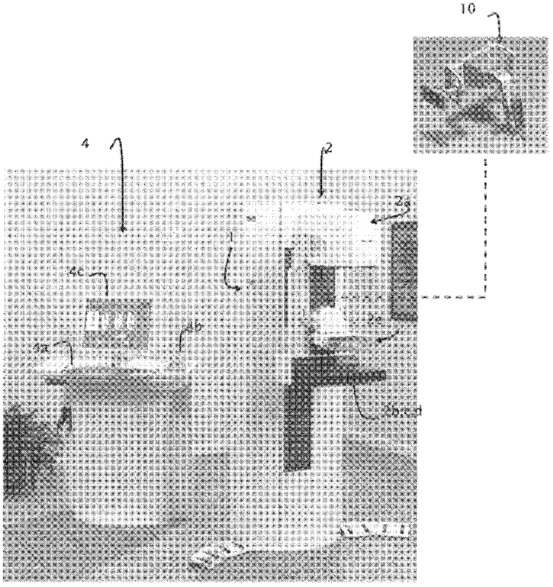
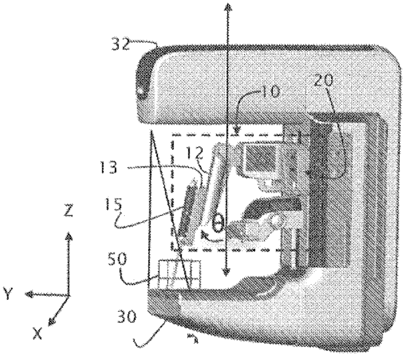
[0025] A breast tomosynthesis system generally includes an X-ray source mounted on a rotating arm of a gantry and an X-ray detector, the X-ray detector being usually positioned so that when the X-ray detector In a zero position, the X-ray detector is approximately perpendicular to the X-ray source. When acquiring tomosynthetic images, the X-ray source is rotated within a limited angular range. At various points in the path of the X-ray source, the X-ray source is activated and images can be captured by the detector. Each image captured at each point is called a projection image. A computer program is used to reconstruct a three dimensional volume from the projection image, and the three dimensional volume is used for lesion detection. figure 1 One embodiment of an x-ray imaging system that can be adapted to incorporate the present invention is shown in , which can perform mammography and tomosynthesis imaging.

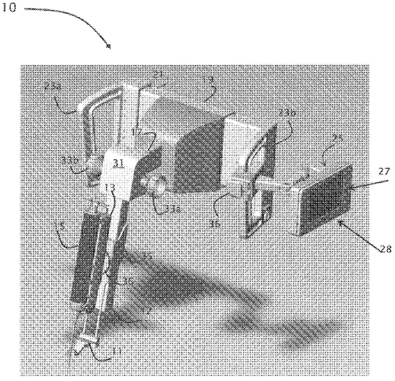
[0026]The shown combined mammography / tomography imaging system...


PUM


Abstract

A tilted needle biopsy assembly is provided for mounting on an x-ray system. Because the biopsy needle is angied relative to at least one of the detector and the x-ray source, x-ray imaging may be performed during the biopsy procedure without interference by the biopsy device. The angled biopsy needle additionally allows improved access to the axilla and chest wall of the patient. The stereotactic biopsy device of the present invention may be coupled to any x-ray system, whether upright or prone, including but not limited to mammography systems, tomosynthesis systems, and combination mammography/tomosynthesis systems. The system flexibly supports the use of any mode of image capture (i.e., scout, two dimensional mammogram, three-dimensional reconstructed volume) for either or both target visualization and target localization. With such an arrangement, a needle biopsy assembly having improved patient coverage is provided for use with a variety of different x-ray imaging platforms.

Description

[0001] related application [0002] Pursuant to Part 35 of the United States Code 1.119(e) (35U.S.C. 1.119(e)), this application claims priority to US Provisional Patent Application Serial No. 61 / 249,772, filed October 8, 2009, which is hereby incorporated by reference. Background technique [0003] Mammography is a well-established method of breast imaging that can be used for screening and diagnosis of breast cancer. Preferably, mammograms are screened annually in female members of the population over the age of 40 or those with breast cancer risk genes. If a mass or calcification (region of interest) is identified on screening mammograms, the patient may request further diagnosis. This diagnosis may include biopsy of the suspicious area and analysis of isolated tissue. [0004] Historically, at the time of breast biopsy, various imaging modalities have been used. Such imaging methods include ultrasound imaging, X-ray imaging and magnetic resonance imaging. Typically, ...

Claims


Application Information

Patent Timeline
IPC IPC(8): A61B10/02A61B10/00
CPCA61B2019/205A61B6/502A61B2017/3409A61B19/201A61B2019/5238A61B6/04A61B6/44A61B10/02A61B90/17A61B2090/376A61B90/11A61B10/0233A61B34/25A61B6/12
Inventor K·德弗雷塔斯I·萧J·拉维奥拉K·皮科特N·A·戈卡纳特斯奥斯A·凡特C·路特
Owner HOLOGIC INC
Why Eureka
Try Eureka