Method and device for measuring or monitoring tissue or cell transmembrane potential changes

A transmembrane potential and cell technology, applied in the field of biomedical engineering, can solve the problems of simple and easy-to-use systems, application limitations, and complex systems, and achieve the goal of improving scientific cognition, wide applicability, and ensuring physiological activity. Effect

Inactive Publication Date: 2014-07-02
XI AN JIAOTONG UNIV
View PDF7 Cites 0 Cited by
  • Summary
  • Abstract
  • Description
  • Claims
  • Application Information

AI Technical Summary

Problems solved by technology

In recent years, some foreign published literature such as Tang D, Li Y, Wong J, etal. "Characteristics of a charged-coupled-device-based optical mapping system for the study of cardiac arrhythmias". J Biomed Opt, 2005, 10 (2):1-7. Using laser and xenon lamp as light source, Brown NH, Dobrovolny HM, Gauthier DJ, etal "A fiber-based ratiometric optical cardiac mapping channel using a diffraction grating and split detector". Biophys j,2007,93 :254-263 use optical fiber and grating for wavelength selection, the system is more complicated
These have failed to realize the ease of use of the system, and the cost is expensive, which limits the application of this technology

Method used

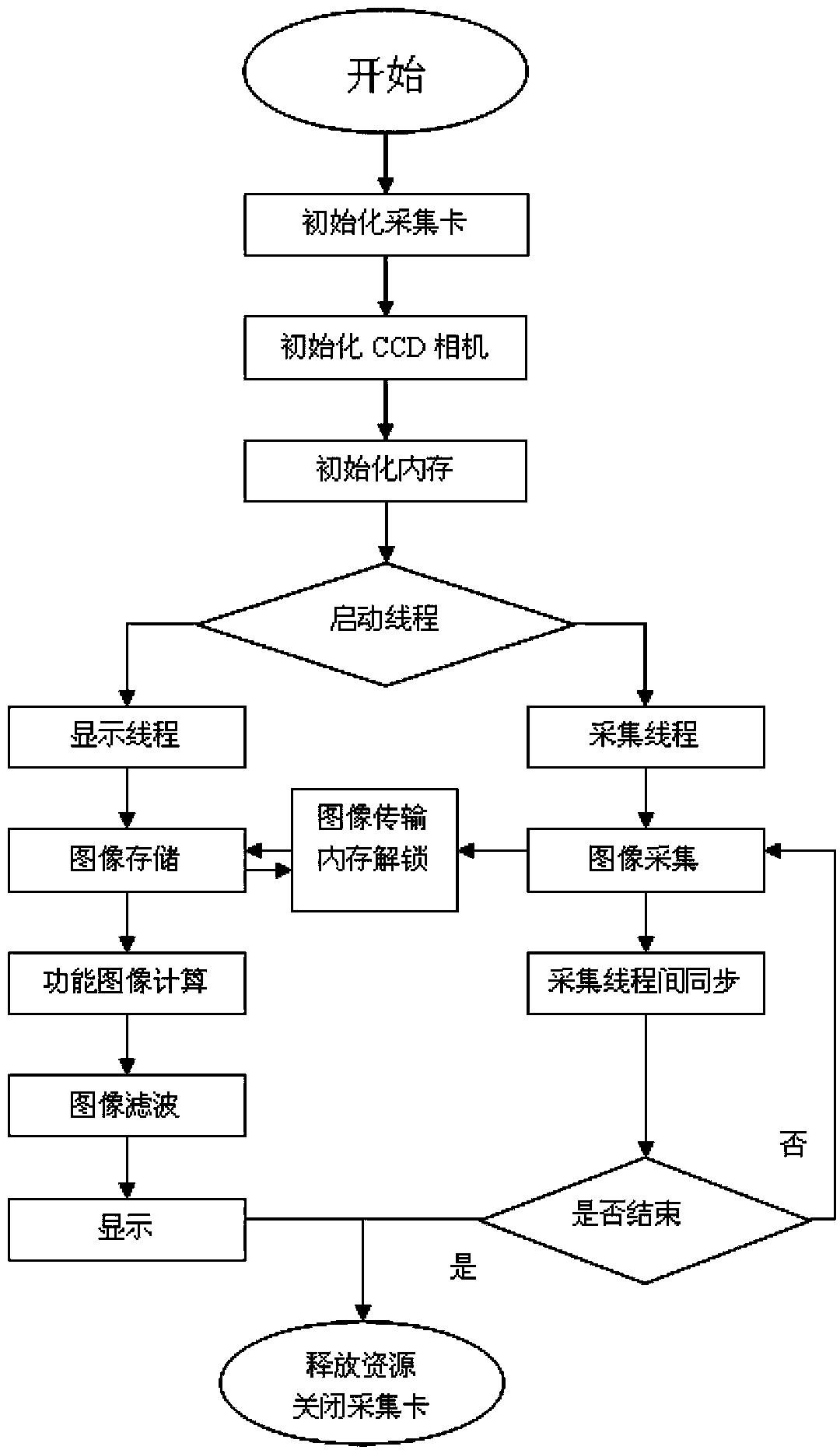

Image

  • Method and device for measuring or monitoring tissue or cell transmembrane potential changes
  • Method and device for measuring or monitoring tissue or cell transmembrane potential changes
  • Method and device for measuring or monitoring tissue or cell transmembrane potential changes

Examples


Embodiment Construction

[0034] The method for measuring or monitoring the change of tissue or cell transmembrane potential in the present invention is as follows: first, the change of cell transmembrane potential is converted into a fluorescent signal, and the change amount ΔF of the light intensity of the fluorescent signal and the change ΔVm of the cell transmembrane potential are detected, according to the following The formula to calculate the optical action potential is:

[0035] V n = - ( F n - F ‾ ) / F ‾

[0036] In the above formula, Vn represents the cell transmembrane potential at a point in the nth frame, Fn represents the fluorescence intensity of the point in the frame, and F represents the average fluorescence intensity of the cell at rest;

[0037]...


PUM


Abstract

The invention discloses a method and a device for measuring or monitoring tissue or cell transmembrane potential changes. The method includes: firstly, converting the cell transmembrane potential changes to fluorescence signals, measuring variable quantity deltaF of the fluorescence signal light intensity and changes deltaVm of cell transmembrane potential, then computing optical action potential, and computing to obtain the action potential for each point so that an equal potential pattern at a certain moment can be reconstructed when the action potential of each point in a frame image is computed; and secondly, utilizing a simplified algorithm to obtain an average resting potential fluorescence image, then directly subtracting a background image from the obtained fluorescence image, and computing to obtain the equal potential pattern finally. The method and the device for measuring or monitoring the tissue or cell transmembrane potential changes can meet requirements of most electrophysiological studies. Dyestuff can be poured onto cell membranes by the aid of a peripheral support system, basic stability is maintained within a measured time, and cell viability is maintained during experiment.

Description

technical field [0001] The invention belongs to the field of biomedical engineering, and relates to a technology that can be used for physiological or pathological cell membrane potential detection. The voltage-sensitive dye is used to convert the membrane potential change signal of cells or tissues into optical information for recording and reconstructing functional images. It is an applied research invented by combining optical technology and image information processing technology in the field of biomedical engineering, especially a method and device for measuring or monitoring tissue or cell transmembrane potential changes. Background technique [0002] The electrical phenomena exhibited by living organisms during life activities are called bioelectricity, and almost all physiological functions are accompanied by some kind of bioelectrical changes. Common detection instruments for these bioelectricity include Near Infrared (NIR), functional Magnetic Resonance Imaging (fM...

Claims


Application Information

Patent Timeline
Patent Type & Authority Patents(China)
IPC IPC(8): G01N27/26
Inventor 张镇西王晶徐正红梅建生隆弢
Owner XI AN JIAOTONG UNIV
Why Eureka
Try Eureka