Fused protein containing cFms extracellular fragments, preparation method and applications of fused protein with cFms extracellular fragments

A technology for fusion proteins and extracellular fragments, applied in the field of fusion proteins and their preparation, can solve problems such as expression differences and incomplete intracellular signal transmission pathways

Inactive Publication Date: 2013-04-17
SUZHOU UNIV
View PDF3 Cites 5 Cited by
  • Summary
  • Abstract
  • Description
  • Claims
  • Application Information

AI Technical Summary

Problems solved by technology

Although there are many biological functions similar to each other, the intracellular signaling pathways used after receptor binding are not completely the same, and there are differences in the expression of each other in tissues

Method used


Image

  • Fused protein containing cFms extracellular fragments, preparation method and applications of fused protein with cFms extracellular fragments
  • Fused protein containing cFms extracellular fragments, preparation method and applications of fused protein with cFms extracellular fragments
  • Fused protein containing cFms extracellular fragments, preparation method and applications of fused protein with cFms extracellular fragments

Examples


Embodiment 1

[0049] Construction of expression plasmids:

[0050] The coding sequence of the extracellular region of cFms was obtained by gene synthesis method according to GenBank (BC047521.1).

[0051] The Fc fragment (684bp) is amplified by PCR with primer 1 and primer 2 using lymph node cDNA (BD) as a template:

[0052] Primer 1: 5'-ctatctcacacatcgacaattcgaagacaaaactcacacatgcccac-3'

[0053] Primer 2: 5'-aagggaatctagagcggccgctcatttacccggagacaggggag-3'

[0054] YY-001 consists of the 1st, 2nd and 3rd immunoglobulin-like regions (D1, D2, D3) of the extracellular region of cFms (see figure 2 ) fused with human immunoglobulin Fc. Between the extracellular domain D3 and Fc, a connecting peptide is added. The obtained DNA sequence of YY-001 is shown in SEQ ID NO.19. Among them, the PCR fragment (831bp) of D1 to D3 is obtained by PCR amplification with primer 3 and primer 4:

[0055] Primer 3: 5'-ccgctcgagatcccagtgatagagcccagt-3'

[0056] Primer 4: 5'-gcataccggttaccacccggaagaacatgga-3...

Embodiment 2

[0073] Transfection of host cells and production of fusion proteins:

[0074] A plurality of fusion proteins in the present invention are expressed in CHO-K1, CHO-S and DG44 cells, secreted into culture fluid, and purified by Staphylococcus A protein affinity precipitation method.

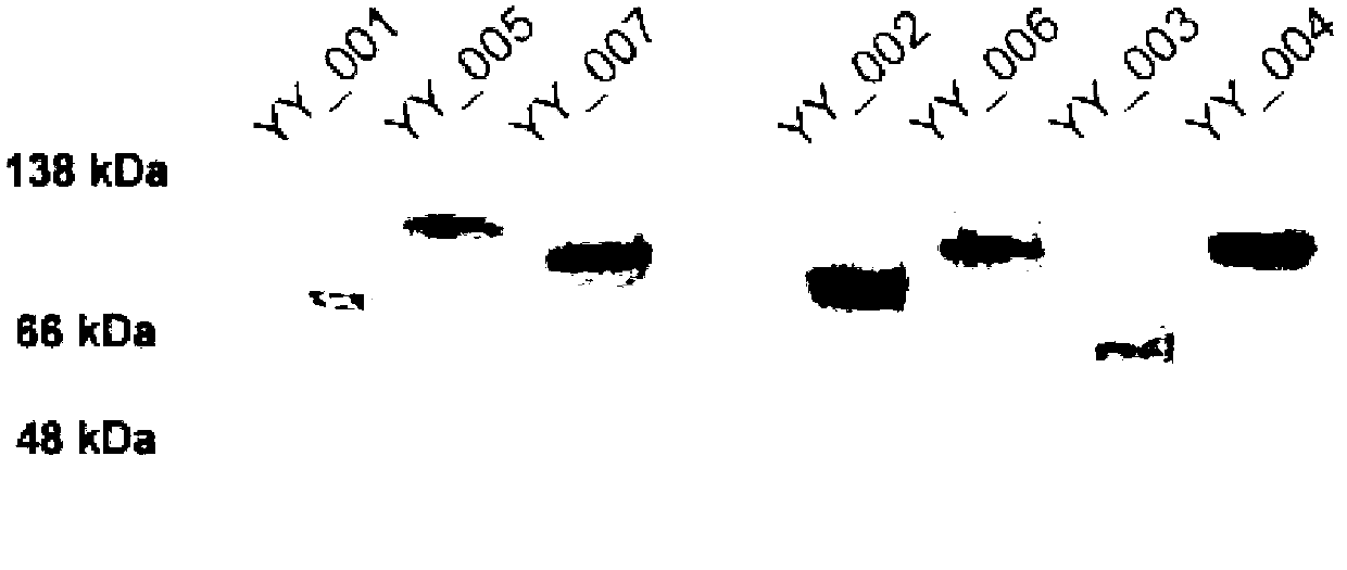
[0075] Transient expression of YY-001 to YY-007 fusion protein in the present invention, the recombinant plasmid was purified with a DNA purification kit (Qiagen Company), and then transfected into CHO-K1 (ATCC#CCL61) cells with Liposome 2000 (Invitrogen Company) . Supernatants were collected after 3 days of culture in serum-free medium OPTI-MII. After the concentration of the purified fusion protein was determined by ELISA, it was verified by SDS-PAGE and western blot analysis.

[0076] The fusion protein of YY-001 to YY-007 in the present invention is stably expressed, and the purified plasmid is transfected into CHO-S cells or DG44 cells (Invitrogen Company) by electroporation. After 48 hours...

Embodiment 3

[0078] In vitro binding experiment of fusion protein with IL-34 and M-CSF:

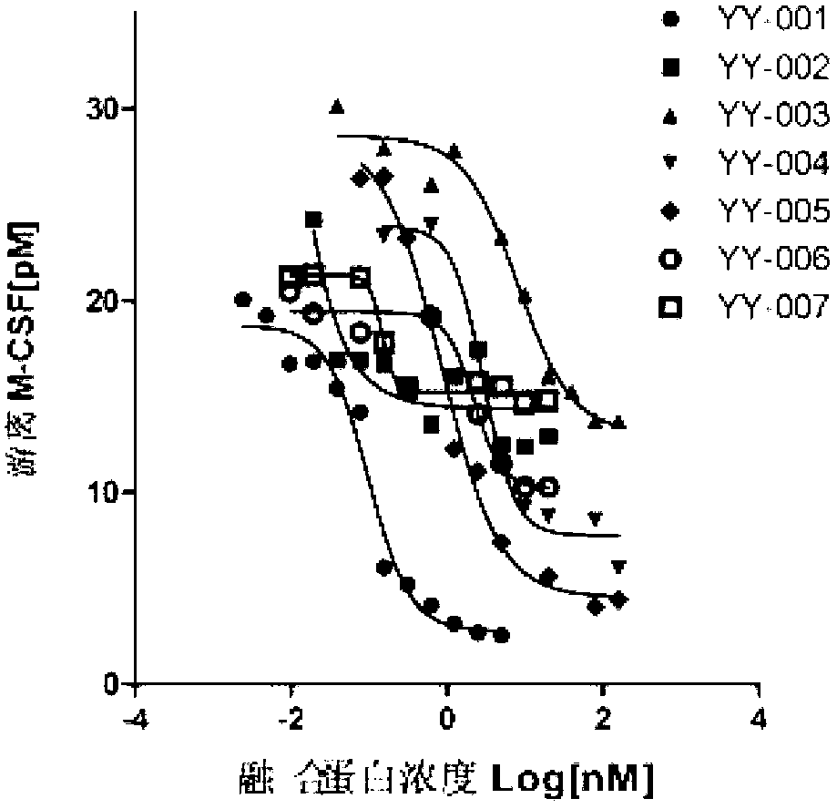
[0079] The present invention uses a highly sensitive and specific IL-34 and M-CSF detection kit (R&D Systems Company) to determine the affinity of each fusion protein to IL-34 and M-CSF. Fusion proteins (YY-001 to YY-007) of different concentrations (0 to 100nM) were incubated with 250pM human IL-34 or with 50pM human M-CSF (R&D Systems) overnight at room temperature, and then treated with IL-34 or The M-CSF detection kit detects free IL-34 and free M-CSF that are not bound by the fusion protein. The experimental results are shown in Table 1.

[0080] Table 1

[0081]

[0082]

[0083] Under the experimental conditions, the fusion proteins YY-001 to YY-007 showed different degrees of affinity to IL-34 or M-CSF. Among them, the affinity of YY-001 to IL-34 and M-CSF is about 0.101-0.149nM / 0.099-0.169nM respectively; the affinity of YY-007 to IL-34 and M-SCSF is about 0.125nM / 0.14nM respectivel...


PUM


Abstract

The invention provides a fused protein with extracellular fragments of a macrophage stimulating factor receptor (cFms). The fused protein is formed by connecting the cFms extracellular fragments and human immunoglobulin Fc by a connecting peptide and is named as cFmsECD-Fc. The cFms extracellular fragments are composed of at least two of five immunoglobulin (IgG) extracellular structure domains. The formed fused protein comprises YY-001, YY-002, YY-003, YY-004, YY-005, YY-006 and YY-007. The fused protein provided by the invention has functions of combining interleukin-34 (IL-34), combining the macrophage stimulating factor receptor (M-CSF) and blocking the combination of IL-34 / M-CSF ligand and common receptor cFms, and is suitable for treating diseases caused by the IL-34 / M-CSF under an abnormal condition. The fused protein is promising in the treatment on autoimmune diseases, inflammatory diseases, osteoporosis and tumor. And the fused protein is used for preparing medicines capable of inhibiting the inflammatory diseases, medicines capable of inhibiting the osteoporosis caused by osteoclast or medicines capable of inhibiting the growth and metastasis of the tumor.

Description

technical field [0001] The invention relates to biomedical engineering technology, in particular to a fusion protein containing cFms extracellular fragments and its preparation method and application. Background technique [0002] Interleukin-34 (IL-34) and macrophage stimulating factor (M-CSF) are cell growth factors. Although there is no similarity in DNA sequence, they share receptor cFms. Although there are many biological functions similar to each other, the intracellular signaling pathways used by the receptors are not completely the same, and there are differences in the expression of each other in tissues. [0003] Interleukin-34 and macrophage-stimulating factor are involved in the survival, proliferation, differentiation and activation of monocyte / macrophage lineage cells. In immune response and inflammation, interleukin-34 and macrophage-stimulating factor bind to their receptors (cFms) to activate monocytes / macrophages, promote cytotoxic function, phagocytosis,...

Claims


Application Information

Patent Timeline
Patent Type & Authority Applications(China)
IPC IPC(8): C07K19/00C12N15/85C12N15/81A61K38/17A61K47/48A61P29/00A61P37/02A61P37/06A61P11/06A61P17/06A61P1/00A61P19/10A61P35/00A61P35/04A61P25/00A61P9/10
Inventor 殷勇郑学初
Owner SUZHOU UNIV
Why Eureka
Try Eureka