Obtaining method and device and medical equipment of optimum m1 value

An acquisition method and the best technology, applied in the field of medical imaging, can solve problems such as complicated process and long time consumption, and achieve the effect of reducing misdiagnosis rate and calculation amount

Inactive Publication Date: 2015-06-17
SHENZHEN INST OF ADVANCED TECH
View PDF3 Cites 0 Cited by
  • Summary
  • Abstract
  • Description
  • Claims
  • Application Information

AI Technical Summary

Problems solved by technology

[0005] The purpose of the embodiment of the present invention is to provide a method for obtaining the optimal m1 value, aiming to solve the problem that the method for obtaining the optimal m1 value in the prior art is complicated and takes a long time

Method used


Image

  • Obtaining method and device and medical equipment of optimum m1 value
  • Obtaining method and device and medical equipment of optimum m1 value
  • Obtaining method and device and medical equipment of optimum m1 value

Examples


Embodiment 1

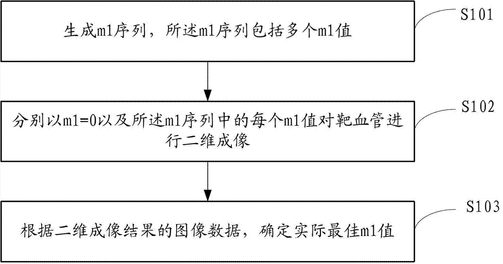
[0038] figure 1 A flow chart showing the implementation of the method for obtaining the optimal m1 value provided by Embodiment 1 of the present invention is described in detail as follows:

[0039] In S101, an m1 sequence is generated, and the m1 sequence includes a plurality of m1 values;

[0040] In S102, perform two-dimensional imaging of the target vessel with m1=0 and each m1 value in the m1 sequence;

[0041] In S103, the actual optimal m1 value is determined according to the image data of the two-dimensional imaging result.

[0042] In this embodiment, by acquiring the m1 sequence, two-dimensional imaging is performed on the target vessel with m1=0 and each value of m1 in the m1 sequence, and the actual optimal m1 is determined according to the image data of the two-dimensional imaging result value. Since only the m1 value is used to perform two-dimensional imaging of the target blood vessel to determine the actual optimal m1 value, the amount of calculation is grea...

Embodiment 2

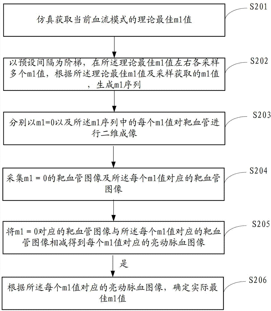
[0044] figure 2 A flow chart showing the implementation of the method for obtaining the optimal m1 value provided by Embodiment 2 of the present invention is described in detail as follows:

[0045] In S201, the simulation obtains the theoretically optimal m1 value of the current blood flow mode;

[0046] In this embodiment, a numerical simulation program is established, and the corresponding relationship between the two-dimensional distribution of the cross-sectional flow velocity in a single voxel and the optimal m1 value is calculated by the numerical simulation program, thereby determining the position of the theoretical optimal m1 value, Since the difference between the theoretical optimal m1 value and the actual optimal m1 value is relatively small, the range of the actual optimal m1 value can be determined by pre-determining the theoretical optimal m1 value, thereby improving the efficiency of obtaining the actual optimal m1 value.

[0047] In S202 , a plurality of m1...

Embodiment 3

[0064] Figure 6 It shows the structural diagram of the device for obtaining the optimal m1 value provided by Embodiment 3 of the present invention. For the convenience of description, only the parts related to the embodiment of the present invention are shown. The device may be a software unit built in medical equipment , a hardware unit or a combination of hardware and software.

[0065] The device for obtaining the optimal m1 value includes: an m1 sequence generating unit 61 , a two-dimensional imaging unit 62 , and an m1 value determining unit 63 .

[0066] m1 sequence generating unit 61, configured to generate m1 sequence, the m1 sequence includes a plurality of m1 values;

[0067] A two-dimensional imaging unit 62, configured to perform two-dimensional imaging of the target vessel with m1=0 and each m1 value in the m1 sequence;

[0068] The m1 value determination unit 63 is configured to determine an actual optimal m1 value according to the image data of the two-dimens...


PUM


Abstract

The invention is applied to the technical field of medical imaging and provides an obtaining method, a device and medical equipment of an optimum m1 value. The method includes generating an m1 sequence, and the m1 sequence is composed of multiple m1 values. Two-dimension imaging on a target vessel is conducted respectively by m1=0 and each m1 value in the m1 sequence. According to image data of a two-dimension imaging result, the optimum m1 value is actually confirmed. Due to the fact that the two-dimension imaging on the target vessel is conducted only by the m1 values and the actual optimum m1 value is confirmed, the method greatly reduces calculated amount, can achieve simple and quick confirmation of the optimum m1 value, ensures obtaining of a high-quality artery image in different patient or parts, reduces misdiagnosis rate, and ensures that a non-enhanced magnetic resonance angiography (MRA) based on a female sexual arousal disorder (FSD) module becomes a vessel imaging manner for clinical use.

Description

technical field [0001] The invention belongs to the technical field of medical imaging, and in particular relates to a method, device and medical equipment for obtaining an optimal m1 value. Background technique [0002] Angiographic imaging technology based on flow-sensitive dephasing (FSD) is a non-contrast-enhanced magnetic resonance angiography method with clinical application prospects in peripheral arteries. This technology utilizes the difference in blood flow velocity between arteries and veins and the sensitivity of the FSD module to blood flow, by subtracting a collection of "black arterial blood" using the FSD module and another collection of "bright arterial blood" without using the FSD module. shadow to obtain the final arterial vessel image. A notable feature of this technology is that the ability of the FSD module to suppress blood flow signals depends on the magnetic field gradient first-order moment (first-order gradient moment, m1) and blood flow velocity....

Claims


Application Information

Patent Timeline
Patent Type & Authority Patents(China)
IPC IPC(8): A61B5/055
Inventor 刘新樊昭阳张娜陈汉威钟耀祖郑海荣李德彪
Owner SHENZHEN INST OF ADVANCED TECH
Why Eureka
Try Eureka