Low-dose X-ray CT image reconstruction method

A CT image and X-ray technology, which is applied in the field of low-dose X-ray CT image reconstruction, can solve the problems of loss of original detail information of the image, failure to meet the real-time imaging requirements of clinical CT, and reduction of CT image resolution.

Active Publication Date: 2013-11-27
SOUTHERN MEDICAL UNIVERSITY
View PDF3 Cites 14 Cited by
  • Summary
  • Abstract
  • Description
  • Claims
  • Application Information

AI Technical Summary

Problems solved by technology

When reconstructing CT images with the same pixel size, the iterative reconstruction method based on the statistical model takes much longer than the classical analytical reconstruction method, so it is far from meeting the needs of clinical CT real-time imaging
[0006

Method used


Image

  • Low-dose X-ray CT image reconstruction method
  • Low-dose X-ray CT image reconstruction method
  • Low-dose X-ray CT image reconstruction method

Examples


Embodiment 1

[0057] A low-dose X-ray CT image reconstruction method includes the following steps in sequence.

[0058] (1) Obtain imaging system parameters of CT equipment and projection data under low-dose CT scanning protocol ; The imaging system parameters of the acquired CT equipment include X-ray incident photon intensity , the variance of the electronic noise of the system .

[0059] (2), according to the projection data in step (1), calculate the projection data variance on each data point ,in represents the location of the data point, Indicates the number of all data points;

[0060] It should be noted that step (2) may use an image-based local variance estimation method or a variance estimation method based on the noise statistical characteristics of CT projection data to calculate the projection data variance on the data points.

[0061] The variance estimation formula can be set according to the needs, for example, the variance estimation formula can be:

[0062] ...

Embodiment 2

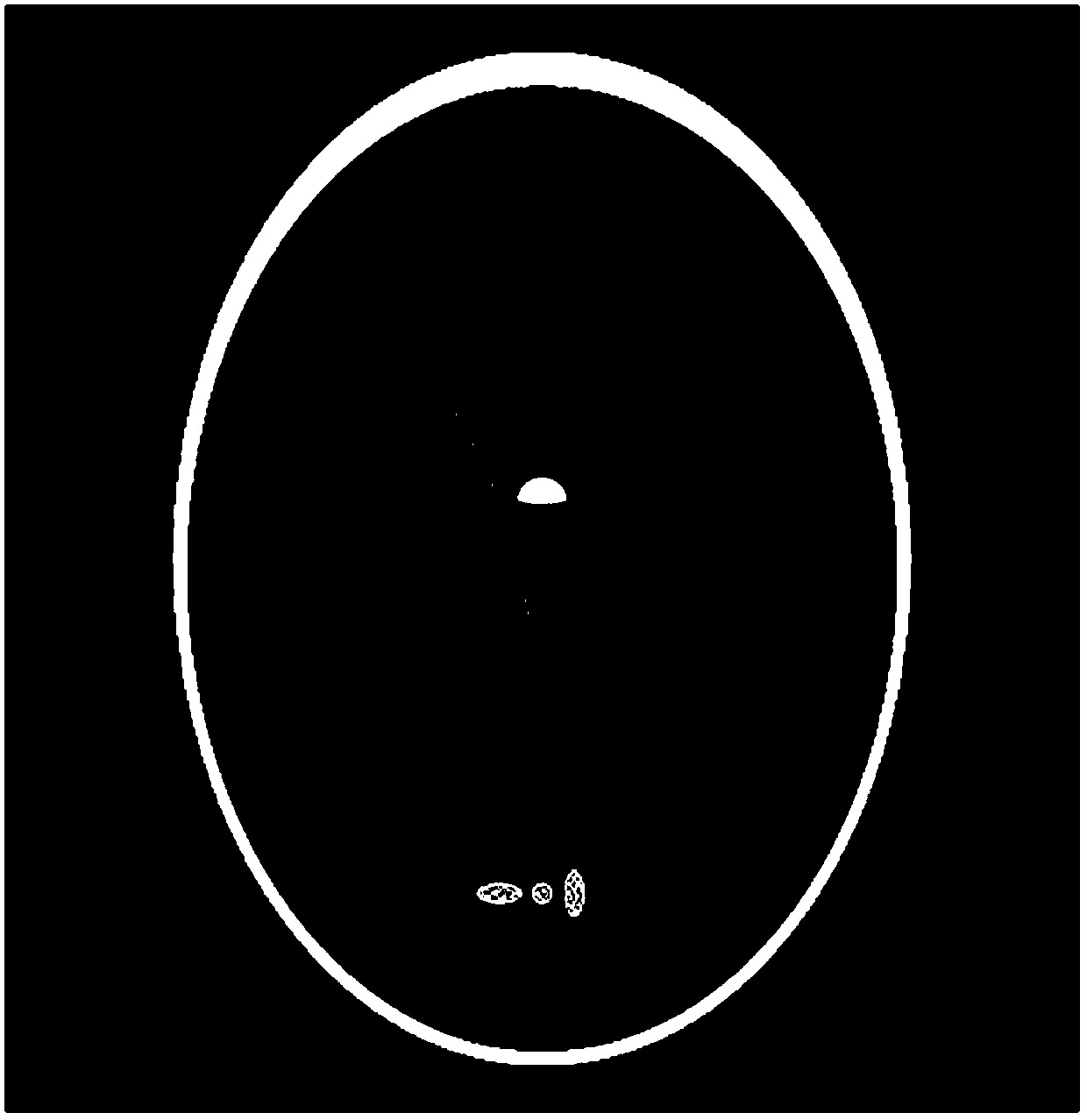
[0097] use as figure 2 The shown Shepp-Logan digital phantom image is used as the computer simulation experiment object of the present invention. The size of the phantom image is set to 512×512, the distances from the X-ray source of the simulated CT equipment to the rotation center and the detector are 1361.2mm and 615.18mm respectively, and the rotation angle is at The interval sampling value is 1160, and each sampling angle corresponds to 672 detector units, and the size of the detector unit is 1.85 mm.

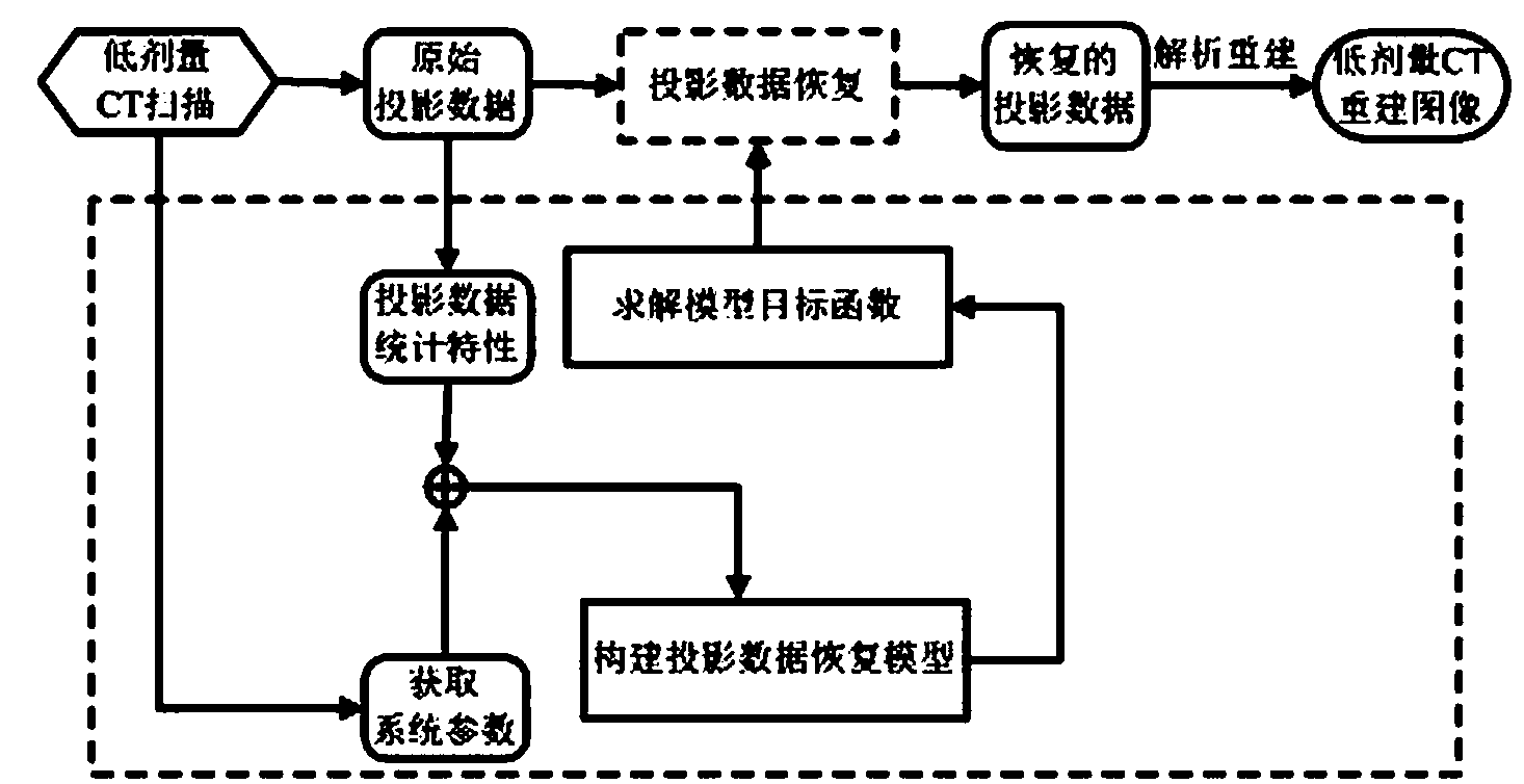
[0098] like figure 1 As shown, the low-dose X-ray CT image reconstruction method of the present invention comprises the following steps in turn:

[0099] (1) Obtain imaging system parameters of CT equipment and projection data under low-dose CT scanning protocol . Imaging system parameters of the obtained CT equipment: incident photon intensity of X-rays 2.5×10 5 , the variance of the electronic noise of the system is 11.0. It should be noted that in actual C...


PUM


Abstract

The invention discloses a low-dose X-ray CT image reconstruction method. The low-dose X-ray CT image reconstruction method sequentially comprises the steps that (1) imaging system parameters of CT equipment and projection data in low-dose CT scanning protocol are obtained, (2) the projection data variance on each data point is calculated according to the projection data obtained in the step (1) and the position of each data point and the number of all the data points are showed, (3) a projection data recovery model is established according to the projection data variances estimated in the step (2), (4) solving is carried out according to the projection data obtained in the step (1) by means of the projection data recovery model established in the step (3) to obtained recovered projection data, and (5) image reconstruction is carried out according to the recovered projection data obtained in the step (4) to obtain a reconstructed CT image. The low-dose X-ray CT image reconstruction method has the advantages that noise and artifact of the image can be greatly reduced, the resolution ratio of the original image can be well kept, and high-quality reconstruction of the CT image is finally achieved.

Description

technical field [0001] The invention relates to the technical field of image processing of medical images, in particular to a low-dose X-ray CT image reconstruction method based on restoration of projection data. Background technique [0002] X-ray CT scanning has been widely used in clinical medical imaging diagnosis, but the excessive X-ray radiation dose in the process of CT scanning has potential risks to the human body, which may easily cause radiation damage and induce malignant tumors. On the premise of ensuring the image quality, minimizing the dose of X-rays has become one of the key technologies in the field of medical CT imaging. [0003] In order to reduce the dose of X-ray radiation, various hardware and software technologies can be used to reduce the dose of X-ray used in CT scanning. Common methods include reducing tube current, reducing X-ray exposure time, and reducing the amount of projection data collected, among which reducing the amount of projection da...

Claims


Application Information

Patent Timeline
IPC IPC(8): G06T5/00
Inventor 马建华牛善洲黄静陈武凡
Owner SOUTHERN MEDICAL UNIVERSITY
Why Eureka
Try Eureka