Medical image segmentation algorithm

A technique for medical images, segmentation algorithms

Inactive Publication Date: 2013-12-04
XIAN HWATECH MEDICAL INFORMATION TECH
View PDF7 Cites 17 Cited by
  • Summary
  • Abstract
  • Description
  • Claims
  • Application Information

AI Technical Summary

Problems solved by technology

The disadvantage of the watershed algorithm is that it requires a continuous "flooding" process, and the amount of calculation is too large for the processing of CT image sequences with a gray value span of 4000 levels and a number of layers of hundreds of layers.
The disadvantage of the super pixel algorithm is that it is easy to over-

Method used


Image

  • Medical image segmentation algorithm
  • Medical image segmentation algorithm
  • Medical image segmentation algorithm

Examples


Embodiment Construction

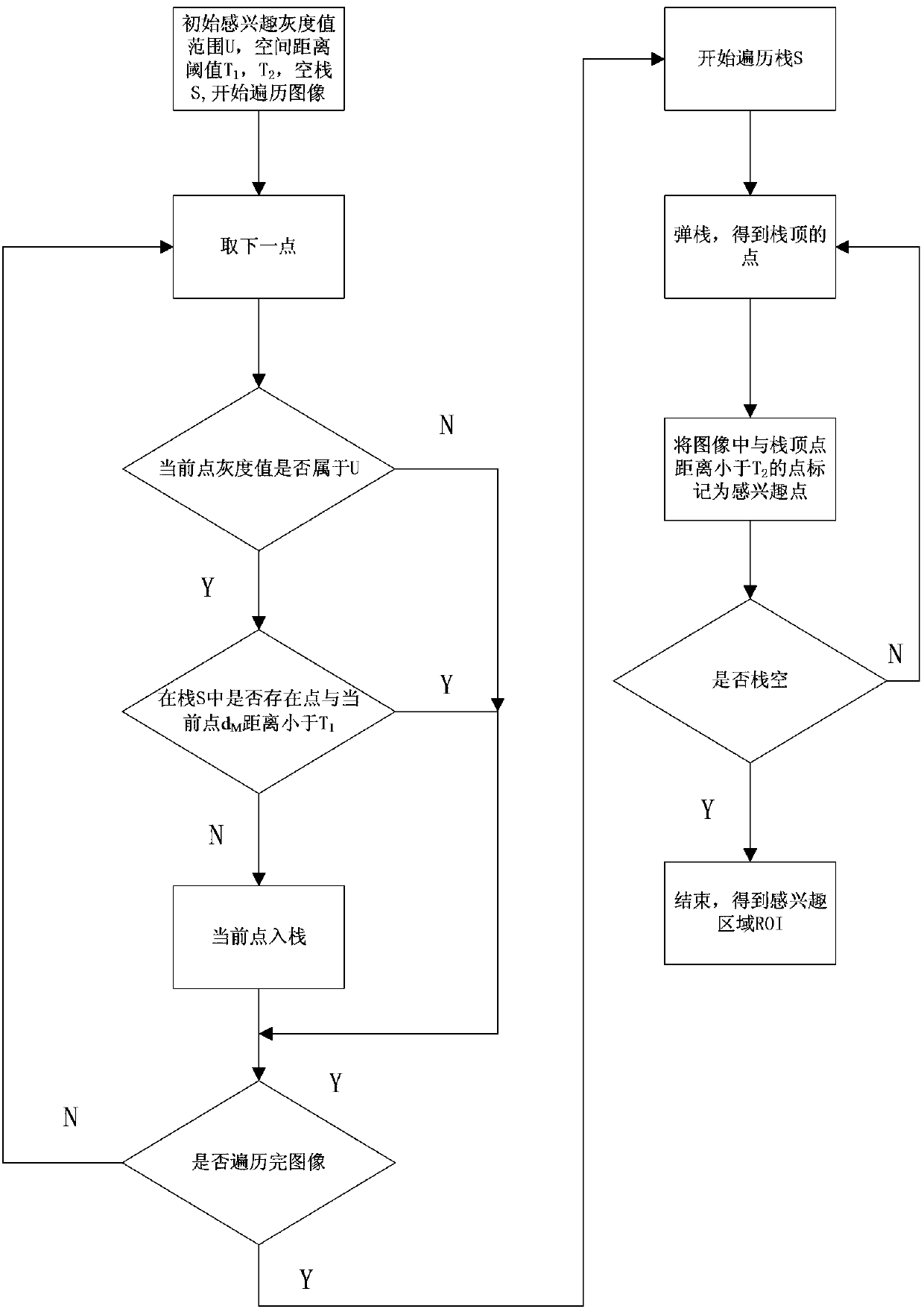
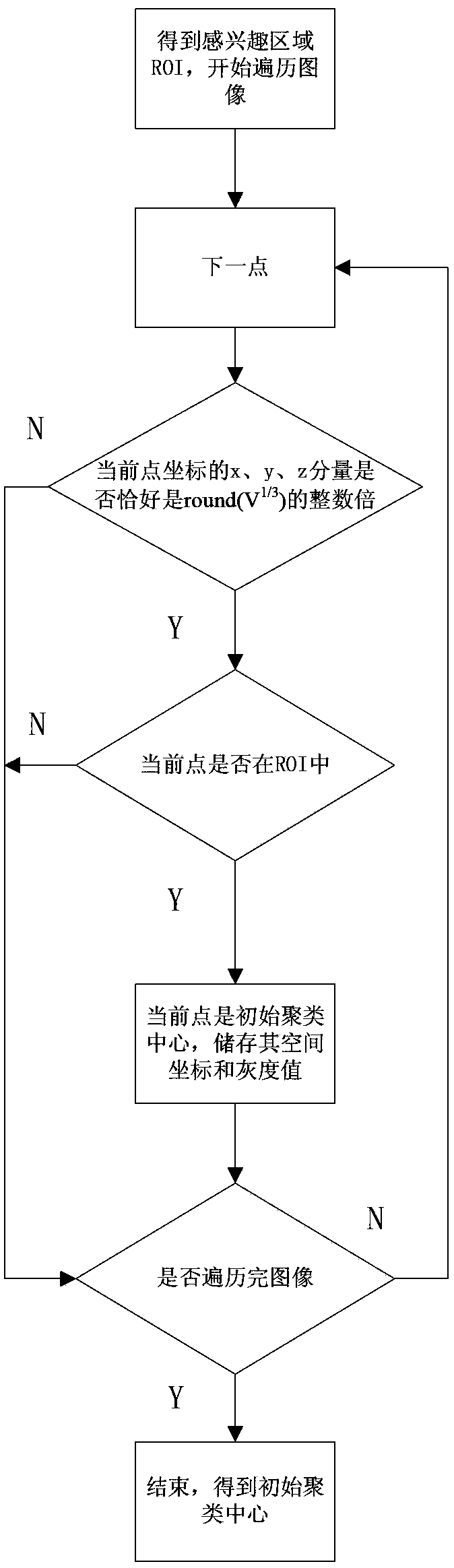
[0040] Such as figure 1 As shown, the medical image segmentation algorithm of the present invention processes a three-dimensional medical image composed of multi-layer two-dimensional images, and the specific steps include selecting a region of interest, arranging initial cluster center points, cluster segmentation and connectivity adjustment.

[0041] Step 1. Select an area of ​​interest

[0042] In practical applications, the image region of interest is often not the entire medical image. Therefore, in order to reduce the calculation time of the algorithm, the region of interest (ROI) of the medical image must first be selected, and the subsequent clustering and segmentation are only performed on the region of interest. . When the ROI is not all medical images, this step removes the segmentation operation on the uninterested regions, which will significantly reduce the running time of the algorithm. Such as figure 2 As shown, step one specifically includes the following ...


PUM


Abstract

The invention discloses a medical image segmentation algorithm and relates to the field of digital image processing technique. The medical image segmentation algorithm includes the steps of selection of an area of interest, arrangement of an initial clustering center, clustering segmentation and connectedness adjustment. In the segmentation algorithm, the area of interest and nearby areas are segmented and divided into a series of homogeneous small areas, follow-up processes can be directly operated at the level of the homogeneous areas instead of single pixel, so that calculated amount is greatly reduced, execution is quickened, and result is accurate. The medical image segmentation algorithm is especially suitable for operations of tissue positioning, measuring, identifying or classifying of a computer controlled X-ray computed-tomography scanning device or a medical radioactive image processing system.

Description

technical field [0001] The invention relates to the technical field of digital image processing, in particular to a medical image segmentation algorithm. Background technique [0002] X-ray computed tomography (Computed tomography, CT) is to use X-rays to scan a layer of a certain thickness of a designated part of the human body. The X-rays that pass through this layer are received by the detector and converted into visible light. The signal is then converted into digital by an analog / digital converter, and stored in the computer as a numerical dot matrix image, which is commonly referred to as a CT image. CT images are an important tool for medical diagnosis. The current technical level cannot realize automatic diagnosis of diseases. Physicians need to diagnose lesions by observing CT images. Under this premise, an effective image processing algorithm can make it easier for doctors to observe and analyze CT images, which can not only speed up the doctor's diagnosis speed, ...

Claims


Application Information

Patent Timeline
IPC IPC(8): G06T7/00A61B6/03
Inventor 王小龙申田李云峰张孝林
Owner XIAN HWATECH MEDICAL INFORMATION TECH
Why Eureka
Try Eureka