
Beta-1 adrenergic receptor monoclonal antibody for inducing myocardial cell apoptosis

A monoclonal antibody and antibody technology, applied in the field of immunology, can solve the problems of high economic cost, not widely used, low concentration of β1-AA, etc., and achieve the effect of improving immunogenicity

Inactive Publication Date: 2014-03-12
CAPITAL UNIVERSITY OF MEDICAL SCIENCES
View PDF2 Cites 3 Cited by
  • Summary
  • Abstract
  • Description
  • Claims
  • Application Information

AI Technical Summary

Problems solved by technology

Through this method, specificity for β 1 -AR-EC II antibody, but get β 1 -The concentration of AA is too low to carry out follow-up experiments, and the economic cost is too high, therefore, this technique is not widely used

Method used


Image

  • Beta-1 adrenergic receptor monoclonal antibody for inducing myocardial cell apoptosis
  • Beta-1 adrenergic receptor monoclonal antibody for inducing myocardial cell apoptosis
  • Beta-1 adrenergic receptor monoclonal antibody for inducing myocardial cell apoptosis

Examples


Embodiment 1

[0026] Embodiment 1: peptide segment synthesis:

[0027] According to people beta 1 The amino acid sequence of the second extracellular loop of the adrenoreceptor was synthesized into two antigenic peptides, wherein the sequence of polypeptide 1 is HWWRAESDEARRCYNDPKCCDFVTNRC; the sequence of polypeptide 2 is CHWWRAES DEARR.

Embodiment 2

[0028] Embodiment 2: Monoclonal antibody is synthesized by the method of hybridoma cell fusion:

[0029] 1) Coupling polypeptide 1 and polypeptide 2 to carrier proteins KLH and BSA respectively as immunogens;

[0030] 2) Using the two immunogens to immunize three mice respectively to prepare multiple antiserum;

[0031] 3) Biological activity verification with multiple antiserum;

[0032] 4) selecting 1-2 mice with the highest titer, and performing fusion respectively to prepare monoclonal hybridoma cell lines;

[0033] 5) Screening the obtained hybridoma cells by ELISA method, selecting 1 to 5 high-affinity specific monoclonal antibody hybridoma cell lines, and expanding and culturing them;

[0034] 6) One selected clone was inoculated into mice, ascites was collected and Protein G was purified.

Embodiment 3

[0035] Embodiment 3: the biological activity test of the obtained monoclonal antibody

[0036] Detection of β by ELISA, western blotting and laser confocal technology 1 -AA monoclonal antibody and cell surface β 1 -A combination of AR.

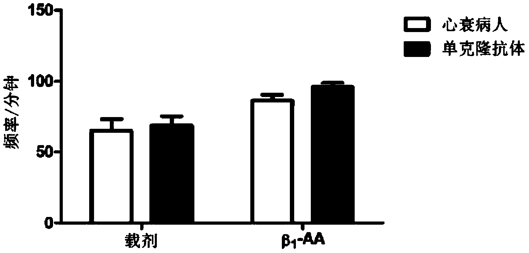
[0037] Experimental evaluation of beating frequency of neonatal rat cardiomyocytesβ 1 - Function of the AA monoclonal antibody.

[0038] Annexin V / PI double staining was detected by flow cytometry β 1 -AA monoclonal antibody against cardiomyocytes H9C 2 Apoptotic effects.

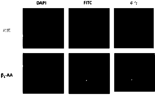
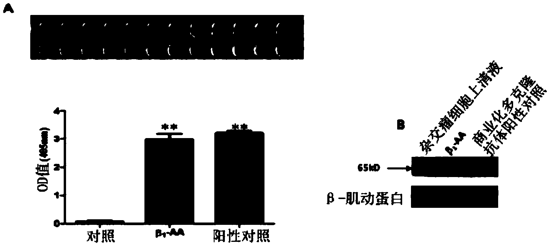
[0039] The results of ELISA detection showed that in the supernatant of hybridoma cells, β 1 The content of -AA was significantly higher than that of the control group (2.9896±0.1873vs.0.0635±0.0386, Pfigure 1 A); and the results of Western blot also confirmed that β 1 -AA monoclonal antibody and commercial anti-β 1 -AR polyclonal antibody positive control has bands with the same molecular weight ( figure 1 B); confocal laser results show that β 1 -AA mAb can inte...


PUM


Abstract

The invention provides a beta-1-AA monoclonal antibody. The monoclonal antibody is produced by using the following two polypeptides as an immunogen, in which polypeptide 1 is a polypeptide with a sequence having 85%-100% identity with HWWRAESDEARRCYNDPKCCDFVTNRC; polypeptide 2 is a polypeptide with a sequence having 85%-100% identity with CHWWRAES DEARR. The beta-1-AA monoclonal antibody is immunized by using two peptide fragments, wherein the polypeptide 1 fragment is an amino acid sequence on a second ring outside a beta-1 adrenergic receptor cell but has low immunogenicity; while the polypeptide 2 fragment is a peptide fragment with high immunogenicity and is designed for the polypeptide 1 fragment. Therefore, the immunogenicity is improved while guaranteeing acquisition of a correct antibody when the monoclonal antibody is immunized by using the two peptide fragments respectively. The monoclonal antibody which aims at the second ring outside the beta-1 adrenergic receptor cell and has the same biological effect as beta-1-AA in the serum of a clinical patient is successfully prepared for the first time. A reliable experiment result can be obtained when the monoclonal antibody is used in scientific research.

Description

technical field [0001] The invention belongs to the field of immunology and relates to a novel β1 adrenergic receptor monoclonal antibody for inducing cardiomyocyte apoptosis. Background technique [0002] beta 1 Adrenoceptor autoantibodies are a beta- 1 Autoantibodies produced by the second extracellular loop of adrenergic receptors are mainly present in various cardiovascular diseases such as dilated cardiomyopathy, Chagas disease and heart failure. The study found that β 1 Adrenergic receptor autoantibodies are of great significance for studying the immunological mechanism of cardiovascular diseases, and the autoantibodies can directly induce cardiomyocyte apoptosis (Daniel Jane-wit et al., Beta1-adrenergic receptor autoantibodies mediate dilated cardiomyopathy by agonistically inducing cardiomyocyte Apoptosis. Circulation. 2007;116:399-410.). Most of the autoantibodies used in previous studies were polyclonal antibodies obtained from serum. The titer of polyclonal a...

Claims


Application Information

Patent Timeline
IPC IPC(8): C07K16/28
Inventor 刘慧荣杜芸辉李笑张苏丽商建宇马新亮
Owner CAPITAL UNIVERSITY OF MEDICAL SCIENCES
Learn More