Tumor metastasis unit counting method based on quantum dot spectrum analysis and image parsing

A technology for tumor metastasis and spectral analysis, which is applied in the field of tumor metastasis unit counting based on quantum dot spectral analysis and image analysis, which can solve the problems of impossible manual calculation, large individual differences, and low resolution, so as to avoid individual judgment errors. , The effect of fast detection speed and simple operation steps

Inactive Publication Date: 2014-04-30
李雁
View PDF7 Cites 5 Cited by
  • Summary
  • Abstract
  • Description
  • Claims
  • Application Information

AI Technical Summary

Problems solved by technology

[0003] In addition, the current calculation of tumor metastatic units is based on immunohistochemical multicolor staining method, the resolution between different colors is low, the error is large, and the manual counting method is used, and individual differences are large, especially in the number of tumor metastatic units. When it is huge, it is almost impossible to complete manual calculations, which seriously hinders its development in medical clinical applications

Method used


Image

  • Tumor metastasis unit counting method based on quantum dot spectrum analysis and image parsing
  • Tumor metastasis unit counting method based on quantum dot spectrum analysis and image parsing
  • Tumor metastasis unit counting method based on quantum dot spectrum analysis and image parsing

Examples


Embodiment 1

[0029] A. Paraffin section of tumor tissue: paraffin-embedded tumor tissue section after formalin fixation, with a thickness of 4 μm, fixed on a detachment-proof glass slide treated with polylysine;

[0030] B. Preparation of tumor tissue slices: Bake the tissue slices in an oven at 60°C for 2 hours, then dewax xylene for 3 times (different dewaxing cylinders), 5 minutes each time, put the tissue slices in turn after dewaxing Absolute ethanol for 5 minutes, 95% alcohol for 2 minutes, 90% alcohol for 2 minutes, 85% alcohol for 2 minutes, and running water for 3 minutes;

[0031] C. Antigen retrieval of tissue sections: prepare trisodium citrate buffer with 29.41 g of trisodium citrate and 1000 ml of double distilled water, prepare citric acid buffer with 10.5 g of citric acid and 500 ml of double distilled water, and take trisodium citrate buffer respectively 20.25ml of citric acid buffer solution, 4.75ml of citric acid buffer solution and 225ml of double distilled water were a...

Embodiment 2

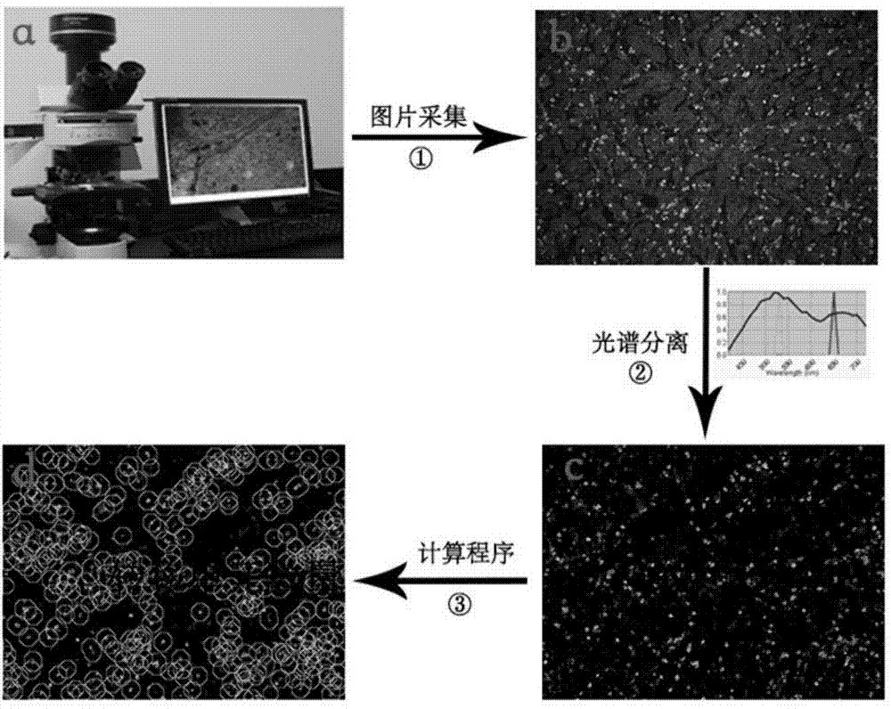
[0041] Image acquisition and counting of tumor metastatic units are as follows:

[0042] 1) The CRi Nuance multi-spectral cell imaging system (Cambridge Research&Instrumentation, Inc., Woburn, MA, USA), excited with ultraviolet light, collected quantum dot double-stained images of tumor tissue ( figure 1 b); The green macrophage signal and the red tumor neovascularization signal were isolated in the tumor tissue area ( figure 1 c);

[0043] 2) The number of tumor metastases is centered on the green signal and expanded around with a radius of 30 microns. If a red signal appears within this range, it is counted as 1, otherwise it is 0, and the number of tumor metastases in this picture is finally output. details as follows:

[0044] a) Image preprocessing: Due to the influence of noise on tumor tissue quantum dot double-stained images, there are a large number of pseudo-minimum points, which easily lead to serious over-segmentation of the watershed. Before the segmentation is...

Embodiment 3

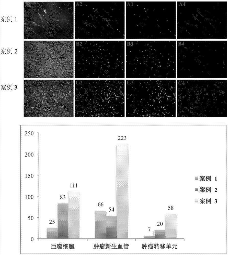
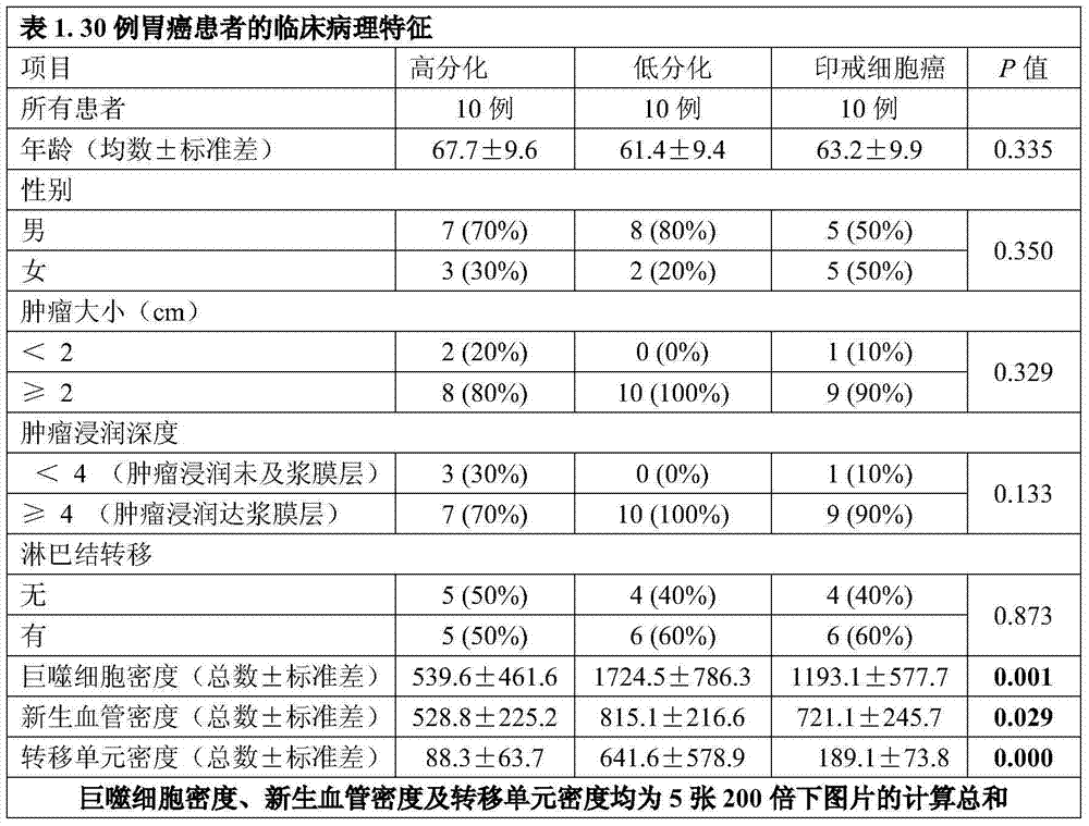
[0051] Validity of tumor metastatic unit counts:

[0052] The applicant counted tumor metastatic units in 30 patients with gastric cancer, including 10 cases of well-differentiated tubular adenocarcinoma, 10 cases of poorly differentiated tubular adenocarcinoma, and 10 cases of signet ring cell carcinoma. There was no difference in the basic clinical parameters, but a single macrophage, The counts of neovascularization and tumor metastasis units were statistically different among the three groups, which confirmed the validity of calculation and analysis of tumor metastasis units.

[0053]


PUM

PropertyMeasurementUnit
thicknessaaaaaaaaaa
radiusaaaaaaaaaa

Abstract

The invention discloses a tumor metastasis unit counting method based on quantum dot spectrum analysis and image parsing, which can be used for rapidly realizing the in-situ imaging of a plurality of components in a tumor microenvironment, the quantitative calculation of each component and the quantitative counting of tumor metastasis units. According to the method, the shortcomings of conventional and modern methods are overcome; the method is high in detection speed and stability; manual calculation is eliminated, so that individual judgment errors are avoided; a metastasis unit consisting of the components can be effectively, conveniently, rapidly and quantitatively calculated, and the development of individualized tumor treatment is favorably promoted.

Description

technical field [0001] The invention belongs to the field of tissue immunofluorescence and image spatial relationship analysis, and specifically relates to a method for counting tumor metastasis units based on quantum dot spectrum analysis and image analysis, which is suitable for tumor cells, macrophages, and neoplastic tumors in the microenvironment of human tumor tissues. The counting of tumor metastatic units composed of three components of blood vessels (vascular endothelial cells) also involves quantitative calculations to detect units composed of other important microenvironmental components. Background technique [0002] Cancer is the leading cause of death in the world. In the past 20 years, its incidence and mortality have remained high, seriously threatening the health of Chinese residents (Jemal A, Bray F, Center MM, et al. Global cancer statistics. CA: A Cancer Journal for Clinicians. 2011, 61:69-90.). The occurrence and development of cancer is a process of mu...

Claims


Application Information

Patent Timeline
Patent Type & Authority Applications(China)
IPC IPC(8): G01N21/64
Inventor 方敏李雁屈爱平刘娟袁静萍
Owner 李雁
Why Eureka
Try Eureka