
Fluorescent quantitative primer group for visual differential diagnosis of waterfowl parvoviruses

A waterfowl parvovirus and differential diagnosis technology, applied in the direction of microorganism-based methods, microorganism measurement/testing, microorganisms, etc., to achieve high efficiency and accuracy, and simple identification methods

Active Publication Date: 2014-06-25
INST OF ANIMAL HUSBANDRY & VETERINARY FUJIAN ACADEMY OF AGRI SCI
View PDF0 Cites 6 Cited by
  • Summary
  • Abstract
  • Description
  • Claims
  • Application Information

AI Technical Summary

Problems solved by technology

[0007] At present, there are no relevant research reports at home and abroad that only need a set of real-time fluorescent quantitative PCR primers to simultaneously perform visual differential diagnosis of GPV and MDPV infection. The establishment of the present invention can fill in the gaps in related fields at home and abroad

Method used


Image

  • Fluorescent quantitative primer group for visual differential diagnosis of waterfowl parvoviruses

Examples


Embodiment 1

[0029] 1. Poison:

[0030] Both goose fine viruses and Pan duck fine viruses are separated and preserved by the Institute of Animal Husbandry and Veterinary Medicine of the Fujian Academy of Agricultural Sciences.

[0031] 2. Primer design and synthesis

[0032] According to the GPV and MDPV non -structural protein genes, the real -time fluorescence quantitative PCR primer P1 and P2 are designed.

[0033] Upstream primer P1: 5’ -TTCTTTTGCTCTCTGGGAAAATA -3 ',

[0034] Downstream primers p2: 5’ -gcttttcaatgcc -3 '.

[0035] 3. Real -time fluorescent quantitative PCR amplification

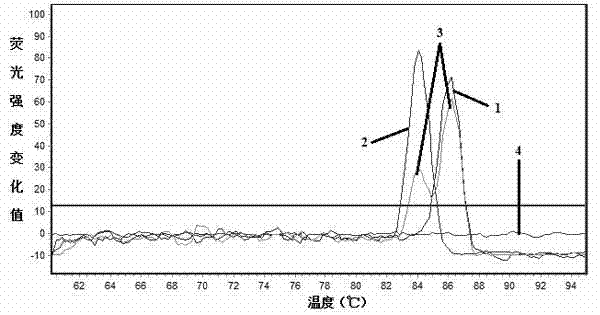
[0036] Extract GPV and MDPV genome DNA in a conventional method.Real -time fluorescence quantitative PCR amplification is used with specific real -time fluorescent quantitative PCR primers P1 and P2.

[0037] Optimized 20 μL of the best reaction system as a system: SYBR Premix EX TAQ TM 10 μL, upstream, downstream primers (10 μmol / L) each 0.2 μL, template 2 μL, and water to make up to 20 μL.The best react...


PUM


Abstract

The invention discloses a real-time fluorescent quantitative PCR (Polymerase Chain Reaction) primer group for visual detection on infection conditions of goose parvovirus (GPV) and muscovy duck parvovirus (MDPV) and a method thereof. According to the method, the detection on the infection conditions of GPV and MDPV is carried out by using dissolution curve temperature difference caused by the difference between nucleotide GC contents of GPV and MDPV specific gene fragment regions amplified by primers, and the infection conditions of GPV and MDPV can be subjected to visual differential diagnosis specifically by only combining the SYBR Green I based real-time fluorescent quantitative PCR primer group to dissolution curves which are automatically generated after reaction is ended. The method disclosed by the invention is simple and is relatively high in efficiency and accuracy.

Description

Technical field [0001] The present invention is an original field of animal molecular disease, which specifically involves a set of fluorescent quantitative primers for the visual diagnosis of small viruses for water poultry. Background technique [0002] Goose Parvovirus (GPV) is the main autonomous replication of small viruses infected with poultry. The scholar Fang Fang was discovered in Yangzhou, Jiangsu Province in 1956 (the introduction of Fang Yi. Little Goose Plague [J].Chinese Veterinary Magazine, 1962, 8: 19-20.], In the future, it is also separated from the virus in many countries. ’ S disease (Wan Chunhe, Zhu Haixia, Huang Yu, waiting for a. A goose fine virus whole genetertex analysis [J]. Journal of Chinese animal infectious diseases, 2011,1,9 (4): 19-24. [0003] Small virus is a small virus department and a small virus, with only one serum type.The disease mainly invaded the fascin and ducks within 30D, causing acute enteritis and liver, kidney, heart and other su...

Claims


Application Information

Patent Timeline
IPC IPC(8): C12Q1/70C12Q1/68C12N15/11C12R1/93
CPCC12Q1/686C12Q1/70C12Q2561/113C12Q2527/107C12Q2563/107
Inventor 万春和黄瑜陈红梅施少华傅秋玲傅光华程龙飞
Owner INST OF ANIMAL HUSBANDRY & VETERINARY FUJIAN ACADEMY OF AGRI SCI
Learn More