Multi-spectral meibomian gland detection analysis device

An analysis device, meibomian gland technology, applied in diagnostic recording/measurement, eye testing equipment, medical science, etc., can solve problems such as poor image contrast, time-consuming and laborious, blurred boundaries between glands and surrounding tissues, and achieve uniform brightness , improve quality, and reduce the effect of local reflections

Inactive Publication Date: 2014-07-16
XIAMEN UNIV
View PDF10 Cites 7 Cited by
  • Summary
  • Abstract
  • Description
  • Claims
  • Application Information

AI Technical Summary

Problems solved by technology

Since the imaging of the meibomian glands needs to penetrate the conjunctiva and epithelial cells, the contrast of the obtained images is poor, and the boundaries between the glands and the surrounding tissues are blurred, and the method of manual observation and counting is time-consuming and laborious

Method used


Image

  • Multi-spectral meibomian gland detection analysis device
  • Multi-spectral meibomian gland detection analysis device
  • Multi-spectral meibomian gland detection analysis device

Examples


Embodiment Construction

[0023] The present invention will be further described below through specific embodiments.

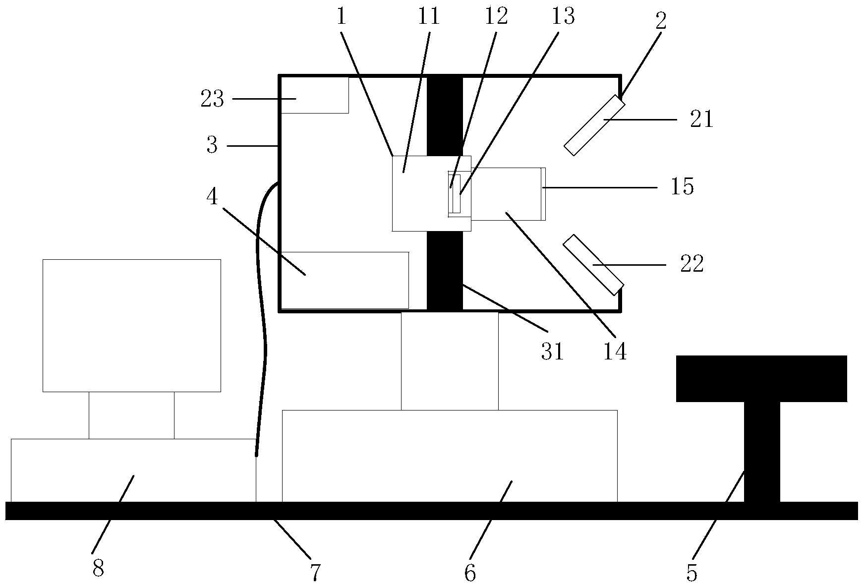
[0024] Such as figure 1 As shown, a multi-spectral meibomian gland detection and analysis device is provided with a multi-spectral imaging system 1, a near-infrared lighting system 2, a three-dimensional motion table 6, a motion controller 4, a tray 5, a housing 3, a base 7 and a host computer 8.

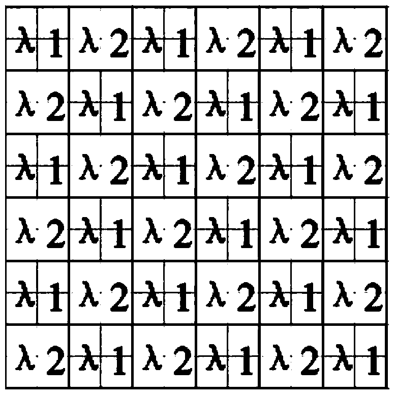
[0025] Such as figure 1 and figure 2 As shown, the multispectral imaging system 1 includes an industrial camera 11 , a narrow-band dual-channel micro-filter 13 , a lens 14 , and a polarizer 15 . The industrial camera 11 is a black and white CCD industrial camera, the number of pixels of the image sensor 12 is 1280(H)*720(V), and the size of the pixel unit is 6.5μm*6.5μm. The narrow-band dual-channel micro-filter 13 is made by micro-lithography technology and vacuum multi-layer coating technology. It includes two narrow-band micro-filter units with different light-passing wavelengths λ1=...


PUM


Abstract

The invention relates to a multi-spectral meibomian gland detection analysis device. The multi-spectral meibomian gland detection analysis device is provided with a multi-spectral imaging system, a near-infrared illumination system, a three-dimensional motion table, a movement controller, a tray, a shell, a base and an upper computer. A narrow-band two-channel micro-optical-filtering sheet is arranged in the multi-spectral imaging system. Two meibomian gland narrow-band spectrum images corresponding to different wave lengths are obtained at the same time through one-time exposure. Pixels of the two images are automatically aligned. Backgrounds of the meibomian gland images can be eliminated by upper computer software through the image fusion algorithm, the meibomian glands are separated from the surrounding tissues, the conditions of the glands can be conveniently observed by a doctor, the indexes such as the number of the glands, the loss rate of the tail ends of the glands, the loss areas of the glands, the opening displacement of the glands and the blocking rate of the gland openings are automatically calculated from the meibomian gland images without the backgrounds, and an objective diagnostic basis is provided for diagnosis of the meibomian glands.

Description

technical field [0001] The invention relates to a medical optical detection device, in particular to an ophthalmic optical detection device for detection and analysis of meibomian glands. Background technique [0002] The meibomian gland is a special differentiated sebaceous gland located in the tarsus of the eyelid, arranged vertically and opening on the eyelid margin, and the lipid secretion discharged is evenly coated on the surface of the tear film through the blinking movement of the eyelid to form the tear film The outermost layer plays an important role in maintaining the health of the ocular surface: it can effectively delay the evaporation of the tear film water layer, reduce the surface tension of the tear film, enhance the stability of the tear film, prevent the skin of the eyelid margin from being soaked by tears, and provide smooth and smooth optics. Interface to reduce damage from blinking and act as a barrier to prevent contamination of the tear film with seba...

Claims


Application Information

Patent Timeline
Patent Type & Authority Applications(China)
IPC IPC(8): A61B5/00A61B3/10
Inventor 张陈涛陈延平刘祖国肖云生颜黄苹卓勇刘婷婷张建寰
Owner XIAMEN UNIV
Why Eureka
Try Eureka