Method to compute and present brain amyloid in gray matter

A technology of gray matter and brain imaging, applied in computing, radiation measurement, computerized tomography scanners, etc., can solve the problem that signals are divided into white matter and gray matter along the projection line

Inactive Publication Date: 2014-07-23
KONINKLJIJKE PHILIPS NV
View PDF4 Cites 2 Cited by
  • Summary
  • Abstract
  • Description
  • Claims
  • Application Information

AI Technical Summary

Problems solved by technology

Additionally, for images where uptake is high in both white and gray matter, it is difficult to separate the signal along the projected line into white and gray matter

Method used


Image

  • Method to compute and present brain amyloid in gray matter
  • Method to compute and present brain amyloid in gray matter
  • Method to compute and present brain amyloid in gray matter

Examples


Embodiment Construction

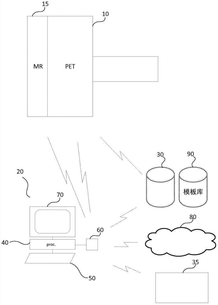
[0017] refer to figure 1 , diagrammatically illustrates an embodiment of an imaging system. The imaging system includes a nuclear scanner 10, such as a PET or SPECT scanner, which scans the subject's brain for the presence or uptake of a radioactive tracer. The scanner records events that occur as a result of the radiotracer emission. The events are recorded in an event list which is transmitted to the imaging workstation 20 which reconstructs an image depicting the distribution of the radiotracer in the brain. Workstation 20 communicates with scanner 10 and may be local or remote. The PET scanner may optionally be combined in other embodiments with other scanners, such as a Magnetic Resonance Imaging (MRI) scanner 15 , that generate images that differentiate between gray and white matter. In one embodiment, the nuclear scanner is combined with an MR scanner and has a common imaging field such that their images are inherently registered.

[0018] Workstation 20 receives im...


PUM


Abstract

An imaging work station (20) includes one or more processors programmed to receive (170)an image depicting a distribution of a radiotracer in a brain or other region of interest. The radiotracer includes at least one of [18F]-Flutemetamol, [18F]-Florbetaben, and [18F]-Florbetapir which highlights amyloid deposits. The image and a template or an MRI image of the region of interest which includes a segmented anatomical feature, such as gray matter, are registered (180)to a common space. A volume representation of the image which depicts the distribution of the radiotracer in the segmented gray matter and suppresses the radiotracer outside of the segmented anatomical feature in white matter is extracted (210).

Description

technical field [0001] This application relates to diagnostic imaging techniques. It finds particular application in the detection of amyloid deposits and will be described with particular reference thereto. Background technique [0002] Amyloid deposition (Aβ) in the brain is one of the hallmarks of Alzheimer's disease. Brain scans are often performed using fluorodeoxyglucose (FDG) radiotracers, but FDG radiotracers do not target Aβ. There are new positron emission tomography (PET) radiotracers for the determination of Aβ that are undergoing phase III clinical trials, such as [18F]-Flutemetamol, [18F]-fluoro Betaben ([18F]-Florbetaben) and [18F]-Florbetapir ([18F]-Florbetapir). One approach to Aβ quantification is kinetic modeling of dynamic PET time series with arterial blood sampling. Although this provides good quantification, clinical application is problematic. A more realistic option uses a comparison of the uptake in the reference area to the target area. The c...

Claims


Application Information

Patent Timeline
Patent Type & Authority Applications(China)
IPC IPC(8): A61B6/03G01T1/29G06T7/00
CPCA61B6/037A61B6/466A61B6/5247A61B5/055G06T7/0012G06T2207/10088G06T2207/10104G06T2207/30016A61B6/501A61B5/0035A61B5/0042G01R33/481G06T7/0014
Inventor F·O·蒂勒F·文策尔
Owner KONINKLJIJKE PHILIPS NV
Why Eureka
Try Eureka