Method for intravascular ultrasound multi-slice shear wave elastography

An elastography and shear wave technology, applied in ultrasonic/sonic/infrasound image/data processing, ultrasonic/sonic/infrasonic diagnosis, ultrasonic/sonic/infrasonic Permian technology, etc. Probes and other issues to achieve high resolution, promote promotion and application, and strengthen early diagnosis capabilities

Inactive Publication Date: 2014-09-24
SUZHOU INST OF BIOMEDICAL ENG & TECH CHINESE ACADEMY OF SCI
View PDF0 Cites 29 Cited by
  • Summary
  • Abstract
  • Description
  • Claims
  • Application Information

AI Technical Summary

Problems solved by technology

At present, the international experimental equipment using intravascular ultrasound imaging technology mainly includes the Eagle Eye? Platinum Catheter produced and developed by Volcano Company of the

Method used


Image

  • Method for intravascular ultrasound multi-slice shear wave elastography
  • Method for intravascular ultrasound multi-slice shear wave elastography
  • Method for intravascular ultrasound multi-slice shear wave elastography

Examples


Embodiment

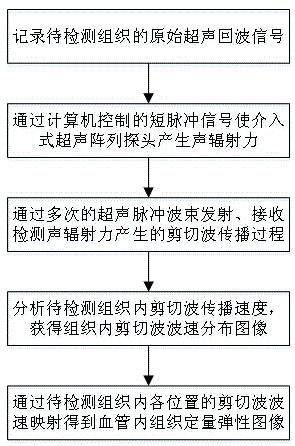
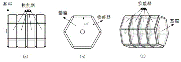
[0031] The intravascular ultrasonic shear wave elastography method of the invention can be used for echo imaging and shear wave elastography, and is suitable for intravascular ultrasonic shear wave elastography of organs and tissues such as the heart. This imaging method needs to insert an imaging transducer into the blood vessel, and the imaging transducer is composed of a plurality of transducer units arranged axially and circumferentially along the base. And each transducer unit is controlled by a multi-channel electronic circuit to emit ultrasonic pulse beams and receive corresponding echo information. The size of each transducer unit and the distance between them can be set according to the shear wave propagation speed in the tissue to be detected. For clarity, in figure 2 , only shows the configuration of a column with a regular hexagonal cross-section as the base, and a total of 24 transducer units of 4×6 arranged on 6 sides. It should be noted that unless changes an...


PUM


Abstract

The invention discloses a method for intravascular ultrasound multi-slice shear wave elastography. The method comprises the following steps of recording an original ultrasound echo signal of a tissue to be detected; enabling an interventional ultrasound array probe to generate acoustic radiation force by virtue of a short-pulse signal controlled by a computer; emitting ultrasonic pulse beams for many times to accept and detect a propagation process of shear waves generated by the acoustic radiation force; analyzing the propagation speed of the shear waves in the tissue to be detected to obtain a wave speed distribution image of the shear waves in the tissue; performing wave speed mapping on the shear waves at each position in the tissue to be detected to obtain a quantitative elastic image of the intravascular tissue. The method has the advantages of real time performance, quantification, high speed, high resolution and the like; a multi-array element intravascular transducer array is used for performing vascular wall tissue elastic information quantitative analysis to excite and track the shear waves; the further popularization and application of intravascular elastography are favorably promoted, and the early diagnosis capability of people in cardiovascular diseases is enhanced.

Description

technical field [0001] The invention relates to an interventional intravascular ultrasonic elastography method, in particular to an elastography method for quickly detecting blood vessel elasticity information in blood vessels by using shear wave elastography technology. Background technique [0002] "China Cardiovascular Disease Report 2012" pointed out that cardiovascular and cerebrovascular diseases have become the number one killer of Chinese people's health in recent years. It is estimated that there are 230 million patients with cardiovascular diseases nationwide, and 3.5 million people die of cardiovascular diseases nationwide every year, accounting for about 41% of all-cause deaths. %, ranking first among all causes of death. The report also predicts that from 2010 to 2030, only considering the factors of population aging and population increase, the number of cardiovascular disease (angina pectoris, myocardial infarction, sudden death of coronary heart disease and s...

Claims


Application Information

Patent Timeline
IPC IPC(8): A61B8/12A61B8/08
CPCA61B8/485
Inventor 崔崤峣焦阳顾天明简小华向永嘉
Owner SUZHOU INST OF BIOMEDICAL ENG & TECH CHINESE ACADEMY OF SCI
Why Eureka
Try Eureka