
Corneal surface protectant as well as preparation method and application thereof

A corneal surface and protective agent technology, applied in the field of medical supplies, to achieve good antibacterial and anti-infection effects, speed up the progress of surgery, and small changes

Active Publication Date: 2014-10-08
JINAN SANWEI MEDICAL INSTR CO LTD
View PDF5 Cites 9 Cited by
  • Summary
  • Abstract
  • Description
  • Claims
  • Application Information

AI Technical Summary

Problems solved by technology

[0007] However, the use of the above-mentioned preservation solution is limited to the preservation of isolated corneal tissue, and has not been used in clinical operations. Whether the above-mentioned preservation solution can be applied to clinical operations, The above-mentioned several technologies are not disclosed, so it is necessary to design a corneal surface protective agent that can be applied in clinical operations, and make the corneal surface protective agent keep the cornea moist and transparent for a period of time, and reduce the midpoint water in the operation. frequency, accelerated surgical progress, and improved patient comfort

Method used


Image

  • Corneal surface protectant as well as preparation method and application thereof
  • Corneal surface protectant as well as preparation method and application thereof
  • Corneal surface protectant as well as preparation method and application thereof

Examples


Embodiment 1

[0061] In cataract extraction + intraocular lens implantation, sodium hyaluronate with a molecular weight of 500,000 is used at a concentration of 9 mg / ml.

[0062] The production method is: weigh 0.045g of NaH 2 PO 4 ·H 2 O, 0.2g of Na 2 HPO 4 , and 0.44g of NaCl, dissolved in 100mL water for injection to prepare buffer solution A, add tobramycin powder to the buffer solution to a final concentration of 3mg / ml, and dexamethasone stock solution to a final concentration of 0.6mg / ml, and stir evenly , until the above-mentioned raw materials are completely dissolved, buffer solution B is obtained, and the buffer solution B is sterilized by filtration with a 0.22 micron filter membrane; 0.9 g of sodium hyaluronate with a molecular weight of 500,000 is dissolved in 100 ml of buffer solution to obtain cornea Protective agent;

[0063] Use a syringe to drop 0.2ml of the product of the present invention on the surface of the cornea, and form a flat liquid interface after 2 second...

Embodiment 2

[0065] Use the method in Example 1 to make a buffer solution, and dissolve 1.5 g of chondroitin sulfate with a molecular weight of 20,000 in 100 ml of buffer solution to obtain a corneal protective agent, which is used in cataract extraction+intraocular lens implantation;

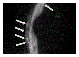
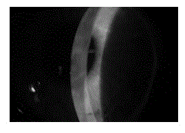
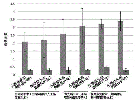
[0066] Use a syringe to drop 0.2ml of the product of the present invention on the surface of the cornea. After 2 seconds, a liquid interface with a flat surface will be formed. The maintained good refractive state and flat surface will last for about 10 minutes. 1 time. A clinical investigation was carried out on the postoperative effects of 16 patients, including 10 in the normal saline group and 6 in the corneal protective agent group. T test was used to compare the statistical differences between the two experimental groups, and P image 3 ) (P Figure 4 ) (P<0.05).

Embodiment 3

[0068] Use the method in embodiment 1 to make buffer, molecular weight is that 700,000 chitosan 1.0g is dissolved in the buffer of 100ml, obtains corneal protectant, is used in cataract extraction+intraocular lens implantation;

[0069] Use a syringe to drop 0.2ml of the product of the present invention on the surface of the cornea. After 2 seconds, a liquid interface with a flat surface will be formed. The maintained good refractive state and flat surface will last for about 10 minutes. 1 time. A clinical investigation was conducted on the postoperative effects of 13 patients, including 5 in the normal saline group and 8 in the corneal protective agent group. T test was used to compare the statistical differences between the two experimental groups, and P image 3 ) (P Figure 4 ) (P<0.05).


PUM

PropertyMeasurementUnit
osmolarityaaaaaaaaaa

Abstract

The invention belongs to the technical field of medical supplies, particularly relates to a corneal surface protectant, and also relates to a preparation method of the corneal surface protectant and an application range of the product. The corneal surface protectant disclosed by the invention comprises the following components: sodium hyaluronate, chondroitin sulfate or chitosan, wherein the molecular weight of the sodium hyaluronate is 100,000-2,000,000D and the concentration range is 5-200mg / ml; the molecular weight of the chondroitin sulfate is 5,000-100,000D and the concentration range is 5-200mg / ml; the molecular weight of the chitosan is 5,000-2,000,000D and the concentration range is 5-200mg / ml. Compared with the traditional preserving fluid, which is just applied to storing in vitro cornea tissue, the corneal surface protectant prepared by the method disclosed by the invention can be applied to a clinical operation, can coat the corneal surface in the operation process, and can keep the wettability and the transmittance of the cornea within a period of time, the dapping frequency in the operation is greatly reduced, the operation schedule is accelerated, and the comfort level of a sufferer is improved.

Description

technical field [0001] The invention belongs to the technical field of medical supplies, and in particular relates to a corneal surface protectant, a preparation method of the corneal surface protectant, and the application range of the product. Background technique [0002] In ophthalmic surgery, keeping the cornea moist and good light transmission is a necessary condition for the operation. In the existing surgery, it is necessary to frequently spot water on the surface of the cornea to achieve the above purpose, while ordinary saline or buffer solution maintains a uniform coverage on the corneal surface. It is difficult to exceed half a minute, and the procedures for dispensing water during the operation are cumbersome, which affects the progress of the operation and increases the discomfort of the patient. The corneal protectant designed in this study is applied to the corneal surface during the operation, which can keep the cornea moist and light-transmitting for a per...

Claims


Application Information

Patent Timeline
Patent Type & Authority Applications(China)
IPC IPC(8): A61K31/737A61P27/02A61P41/00
Inventor 牟国营李志伟
Owner JINAN SANWEI MEDICAL INSTR CO LTD
Learn More