Amphiphilic alpha helix self-assembling peptide and application thereof
A technology selected from, intein, applied in the field of genetic engineering, can solve the problems of restricting the application of inclusion body expression form, complex technology, low yield, etc.
- Summary
- Abstract
- Description
- Claims
- Application Information
AI Technical Summary
Problems solved by technology
Method used
Image
Examples
Embodiment 1
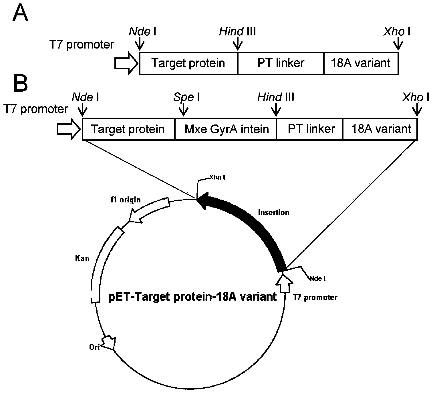
[0065] Example 1: Construction of a fusion protein expression vector using Bacillus subtilis lipase A (LipA) and green fluorescent protein (GFP) as target proteins
[0066] 1.1 Amplification of polynucleotide fragments of 18A variants (19 species)
[0067] Firstly, the nucleotide sequence of the PT-type linker and the 18A variant was designed with the online tool DNAworks. The oligonucleotide primers shown in Table 2 were designed by DNAWorks [13] and synthesized, and then the complete sequence encoding the 18A variant (Hind III-linker-18A variant-Xho I) was obtained by overlapping PCR (overlapping PCR) method. polynucleotide sequence.
[0068] Table 2 is used to amplify the primer list of 18A variant
[0069]
[0070]
[0071]
[0072] a The underlined parts of the primers are the recognition sites of restriction endonucleases Hind III and Xho I, respectively.
[0073] Taking one of the variants, 18Av1, as an example, the specific method for amplifying the polynu...
Embodiment 2
[0093] Example 2: Expression, cell growth status and enzyme activity assay of a fusion protein with Bacillus subtilis lipase A (LipA) as the target protein
[0094] 2.1 Induced expression of fusion protein
[0095] The strain constructed in Example 1 (containing plasmids pET-30a(+)-LipA-native, pET-30a(+)-GFP-18A and pET-30a(+)-GFP-18A variants) was inoculated into the 50 μg / mL kanamycin in LB liquid medium, and cultured in a shaker at 37°C to logarithmic phase (OD 600 =0.4-0.6), add 0.2mM IPTG, induce at 30℃ for 6 hours, harvest the cells, and measure the bacterial concentration OD 600 (The following will be 1mL of OD 600 The amount of cells equal to 1 is called 1OD).
[0096] 2.2 Cell growth status
[0097] The growth state of the cells expressing the LipA-18A variant fusion protein is shown in Table 7 and Figure 4 As shown in A.
[0098] Table 7 expresses the OD of LipA-18A and LipA-18A variant fusion protein cells 600 value
[0099] 18A variant LipA-18...
Embodiment 3
[0107] Example 3: Expression of fusion protein with green fluorescent protein (GFP) as the target protein and its intracellular distribution
[0108] The strain constructed in Example 1 was inoculated into LB liquid medium containing 50 μg / mL kanamycin, and cultivated in a shaker at 37°C to logarithmic phase (OD 600 =0.4-0.6), add 0.2mM IPTG, induce at 23°C for 22 hours, and harvest the cells.
[0109] The harvested cells were treated with 4% paraformaldehyde at 4°C for 1 h. Fluorescent confocal microscopy of GFP cells was performed on a Zeiss710 inverted confocal microscope (Zeiss LSM710confocal microscope) with an excitation wavelength of 488nm.
[0110] The intracellular distribution of active enzyme aggregates induced by 18A and 18A variants was as follows Figure 5 shown. It can be clearly seen from the fluorescent photos that the fluorescence of cells expressing GFP-18A fusion protein is mainly distributed on the inner side of the cell membrane. It shows that the GFP...
PUM
 Abstract
Description
Claims
Application Information
 - R&D
- Intellectual Property
- Life Sciences
- Materials
- Tech Scout
- Unparalleled Data Quality
- Higher Quality Content
- 60% Fewer Hallucinations
Browse by: Latest US Patents, China's latest patents, Technical Efficacy Thesaurus, Application Domain, Technology Topic, Popular Technical Reports.
© 2025 PatSnap. All rights reserved.Legal|Privacy policy|Modern Slavery Act Transparency Statement|Sitemap|About US| Contact US: help@patsnap.com


