Biomarker preserving liquid, biomarker reagent and method

A biomarker and preservation solution technology, which is applied in the preservation of human or animal bodies, biological testing, material inspection products, etc., can solve the problems of poor preservation effect, achieve the effect of convenient use and cost reduction

Inactive Publication Date: 2015-03-25
SHENZHEN MINDRAY BIO MEDICAL ELECTRONICS CO LTD +1
View PDF12 Cites 28 Cited by
  • Summary
  • Abstract
  • Description
  • Claims
  • Application Information

AI Technical Summary

Problems solved by technology

[0016] Some of these preservation solutions are not suitable for the preservat

Method used


Image

  • Biomarker preserving liquid, biomarker reagent and method
  • Biomarker preserving liquid, biomarker reagent and method
  • Biomarker preserving liquid, biomarker reagent and method

Examples


Example Embodiment

[0058] Preparation method of fluorescent marker reagent

[0059] Yet another embodiment of the present invention discloses a method for preparing a fluorescent marker reagent, comprising:

[0060] Provide the above preservation solution and fluorescent tracer-biomarker conjugate;

[0061] Dilute the fluorescent tracer-biomarker conjugate to the working concentration with the preservation solution to obtain a fluorescent marker reagent solution that can be used directly.

[0062] Preferably, the fluorescent tracer-biomarker conjugate is a fluorescently labeled protein, particularly preferably, a fluorescently labeled antibody.

[0063] In the following, a fluorescently labeled protein is taken as an example for illustration.

[0064] Mix fluorescently labeled protein with other components as needed, dissolve in water, and prepare fluorescently labeled protein master solution. Weigh each component of the above storage solution, add water and stir to dissolve, and dilute wit...

Example Embodiment

[0094] Example 1

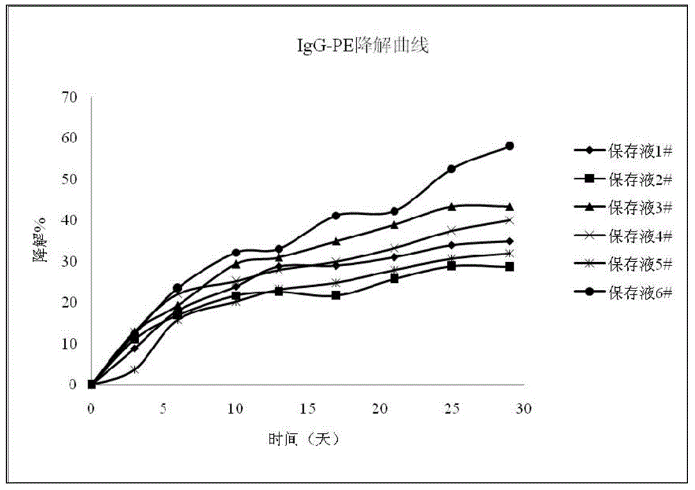
[0095] According to the formula in Table 1, prepare 1#~6# preservation solutions, stir, dissolve and filter at room temperature, and set aside. Prepare self-made fluorescent protein-labeled polyclonal antibody IgG-PE reagents according to conventional methods. Dilute the self-made fluorescent protein-labeled polyclonal antibody IgG-PE reagent with 1~6# preservation solution respectively to a solution of 12.5 μg / mL. The diluted fluorescently labeled polyclonal antibody reagent solution was divided into 9 small portions (50 μL each) and placed in the dark at 40°C. Take a reagent solution every 3 to 4 days and detect the fluorescent tracer according to the aforementioned method- The peak area of ​​the protein conjugate was used to calculate the degradation percentage of the fluorescent tracer-protein conjugate over time, and the curve of time and degradation percentage was drawn.

[0096] Test results such as figure 1 As shown, the degradation percentage o...

Example Embodiment

[0100] Example 2

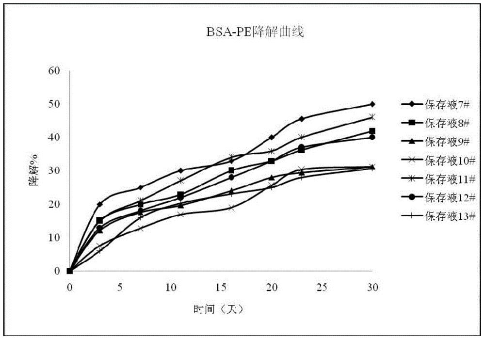
[0101] Prepare 7#~13# preservation solutions according to the formula in Table 2, stir, dissolve and filter at room temperature, and set aside. Fluorescence-labeled protein BSA-PE reagents were prepared according to conventional methods. Dilute the self-made fluorescently labeled protein BSA-PE reagent with 7~13# preservation solution to a solution of 12.5 μg / mL. The diluted fluorescently labeled protein reagent solution was divided into 8 small portions (50 μL each) and placed in the dark at 40°C. Take a portion of the reagent every 3 to 7 days and detect the fluorescent tracer-protein conjugate according to the above method. Calculate the degradation percentage of the fluorescent tracer-protein conjugate with time, and draw the curve of time and degradation percentage.

[0102] Test results such as figure 2 As shown, the degradation percentage of the fluorescent tracer-protein conjugate reflects the stability of the fluorescent tracer-protein conjuga...


PUM


Abstract

The invention discloses a biomarker preserving liquid. The biomarker preserving liquid comprises at least one protein stabilizer, at least one alkaline or neutral amino acid, at least one polyol or polymer, and citrate. The invention also discloses a fluorescent marker reagent prepared by using the preserving liquid, a preparation method of the reagent, and a use of the preserving liquid. The preserving liquid has a very good preserving effect on a fluorescent tracer-biomarker conjugate in the fluorescent marker reagent, can keep the activity and stability of a fluorescent tracer, the biomarker and the conjugate to make the fluorescent marker reagent form a liquid commercial reagent which can be directly used, so the preserving liquid is convenient to use; and the fluorescent marker reagent prepared by using the preserving liquid can tolerate short time high-temperature transportation, so the cost reduction is facilitated.

Description

technical field [0001] The invention relates to the field of in vitro diagnostic reagents, in particular to a storage solution, reagent and method for biomarkers. Background technique [0002] Protein is a macromolecular substance with a complex three-dimensional structure. It is biologically active and susceptible to denaturation under the influence of external conditions. It is easily degraded and inactivated at low concentrations (<0.1mg / mL). It is easy to absorb on the pipe wall and cause loss. For many proteins, typically frozen at -20°C or -80°C, repeated freeze / thaw cycles can denature the protein, causing it to form aggregates with reduced activity. [0003] After the fluorescein is irradiated by the excitation light, it absorbs the light energy and enters the excited state, and can immediately de-excite and emit the emission light (usually the wavelength of the emission light is longer than the wavelength of the incident light, that is, stock shift occurs). At ...

Claims


Application Information

Patent Timeline
IPC IPC(8): G01N33/68G01N33/533
CPCC12N9/96A01N1/0205A01N1/0215G01N33/582G01N33/6809
Inventor 段廷蕊雷霆
Owner SHENZHEN MINDRAY BIO MEDICAL ELECTRONICS CO LTD
Why Eureka
Try Eureka