

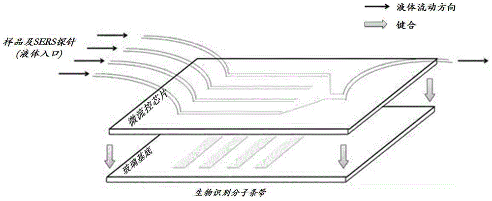
Three-dimensional code biological detection chip based on surface-enhanced Raman scattering (SERS) microflow platform as well as preparation method and detection method of biological detection chip
A biological detection and three-dimensional code technology, applied in the fields of spectroscopy and biological analysis, can solve the problems of complex structure, insufficient information, complicated operation, etc., and achieve the effects of large information capacity, simple preparation and detection methods, and wide application range.
- Summary
- Abstract
- Description
- Claims
- Application Information
AI Technical Summary
Problems solved by technology
Method used
Image

Examples
Embodiment 1
[0029] Example 1: SERS probes were prepared on the basis of gold-core silver-shell nanorods, and high-throughput protein detection was realized by utilizing the specific reaction between antibodies and antigens.
[0030] 1) Preparation of SERS probes.
[0031] To prepare the gold seeds first, mix 2.5 ml of 0.2 M cetyltrimethylammonium bromide (CTAB) solution with 1.5 ml of 1.0 mM tetrachloroauric acid solution at room temperature, stir vigorously and add 0.6 ml of ice-cold 0.01 M hydroboration Sodium solution, stop stirring after 2 minutes to obtain a brown-yellow seed solution. Then prepare a growth solution, add the following reagents in sequence to 50ml 0.2M CTAB solution at room temperature: 2-4ml 4mM silver nitrate solution, 5ml 15mM tetrachloroauric acid solution, 45ml deionized water, and stir slowly evenly. Then 1.5ml-3ml of 0.08M ascorbic acid was added until the solution became colorless. Finally, 1 ml of seed solution was added, and the gold nanorod solution was o...
Embodiment 2
[0040] Example 2: SERS probes were prepared on the basis of gold-core silver-shell nanorods, and high-throughput drug detection was realized by utilizing the specific reaction between antibodies and antigens. :
[0041] 1) Preparation of SERS probes.
[0042] To prepare the gold seeds first, mix 2.5 ml of 0.2 M cetyltrimethylammonium bromide (CTAB) solution with 1.5 ml of 1.0 mM tetrachloroauric acid solution at room temperature, stir vigorously and add 0.6 ml of ice-cold 0.01 M hydroboration Sodium solution, stop stirring after 2 minutes to obtain a brown-yellow seed solution. Then prepare a growth solution, add the following reagents in sequence to 50ml 0.2M CTAB solution at room temperature: 2-4ml 4mM silver nitrate solution, 5ml 15mM tetrachloroauric acid solution, 45ml deionized water, and stir slowly evenly. Then 1.5ml-3ml of 0.08M ascorbic acid was added until the solution became colorless. Finally, 1 ml of seed solution was added, and the gold nanorod solution was o...
PUM
 Abstract
Description
Claims
Application Information