Adipose derived stem cell large-scale culture method
A technology for large-scale culture and quality stem cells, which is applied in the field of large-scale culture of adipose-derived mesenchymal stem cells, which can solve the problems of restricting the application of adipose-derived stem cells, unable to collect cells at one time, and increase the workload, so as to achieve good cell shape and a large number of harvested cells. , the effect of fast cell proliferation
- Summary
- Abstract
- Description
- Claims
- Application Information
AI Technical Summary
Problems solved by technology
Method used
Image
Examples
Embodiment 1
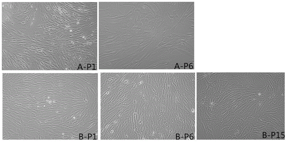
[0051]After primary culture and separation of adipose-derived mesenchymal stem cells confluence reaches 80%, use a pipette to absorb the culture medium in the φ10cm cell culture dish, add 10ml PBS to wash twice, add 2ml TrypLE to digest the adherent cells, and observe under the microscope For cell digestion, after the cells are basically no longer attached to the wall, add 3ml of serum-free medium (UltraCULTURE MEDIUM) to stop digestion, transfer the cell suspension to a centrifuge tube, centrifuge at 1000rpm / min for 5min, remove the supernatant, and use 10ml of serum-free The cells were resuspended in the serum medium, and 20 μl of the cell suspension was taken, and the total number of cells and cell activity information were obtained with a Countstar automatic cell counter. Dilute the cell suspension at 1.3×10 4 / cm 2 Inoculate the cell density into a φ15cm cell culture dish (UltraCULTURE MEDIUM), add 15ml of serum-free medium (UltraCULTURE MEDIUM) containing 10ng / ml EGF, m...
Embodiment 2
[0057] Embodiment 2: cell proliferation curve (traditional method, non-enzyme method)
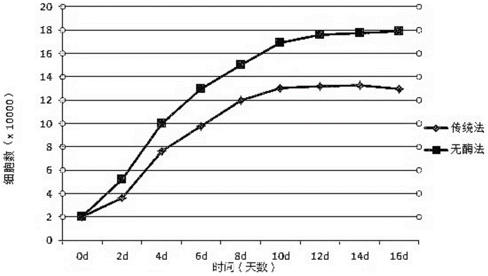
[0058] After the confluence of adipose-derived mesenchymal stem cells separated by primary culture reaches 90%, use a pipette to absorb the culture medium in the φ15cm cell culture dish, add 10ml PBS to wash twice, add 3ml non-enzymatic TrypLE to digest the adherent cells, and mirror Observe the digestion of the cells. After the cells are basically no longer attached to the wall, add 5ml of serum-free medium (UltraCULTURE MEDIUM) to stop the digestion. Transfer the cell suspension to a centrifuge tube, centrifuge at 1000rpm / min for 5min, remove the supernatant, and use Resuspend the cells in 15ml of serum-free medium, take 20μl of the cell suspension, and use the Countstar automatic cell counter to obtain the total number of cells and cell activity information. Dilute the cell suspension at 1.3×10 4 / cm 2 Inoculate the cell density into a φ15cm cell culture dish, add 15ml serum-free mediu...
Embodiment 3
[0060] result: figure 2 The results show that the cell doubling time obtained by the enzyme-free method is significantly better than that of the cells passaged by the traditional method. Example 3: Detection of cell surface markers (P1, P6, P15)
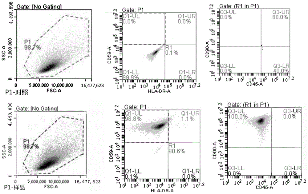
[0061] (1) The culture method of Example 1. Cells were cultured, and the cells of the 1st, 6th, and 15th passages were collected respectively, and the cell surface markers between different passages were detected by flow cytometry. Digest and collect cells separately. After counting, take 4×10⁶ cells from each batch and divide into 4 tubes; wash once with staining buffer, centrifuge at 200 g for 5 min; discard the supernatant, blow and mix the cells with staining buffer; add CD45, CD59, CD90, and HLA-DRA antibodies were 10 μl each, and one tube was used as a blank control; at 4°C, avoid light for 15-20 minutes; wash once with staining buffer, and centrifuge at 1500rpm for 5 minutes; discard the supernatant of directly labeled cells...
PUM
 Abstract
Description
Claims
Application Information
 - R&D
- Intellectual Property
- Life Sciences
- Materials
- Tech Scout
- Unparalleled Data Quality
- Higher Quality Content
- 60% Fewer Hallucinations
Browse by: Latest US Patents, China's latest patents, Technical Efficacy Thesaurus, Application Domain, Technology Topic, Popular Technical Reports.
© 2025 PatSnap. All rights reserved.Legal|Privacy policy|Modern Slavery Act Transparency Statement|Sitemap|About US| Contact US: help@patsnap.com


