

A microscopic analysis device
A microscopic analysis and atomic force microscopy technology, applied in the field of microscopic analysis, can solve the problem that atomic force microscopy imaging cannot qualitatively analyze samples, and achieve the effect of expanding detection methods
- Summary
- Abstract
- Description
- Claims
- Application Information
AI Technical Summary
Problems solved by technology
Method used
Image

Examples
Embodiment 1
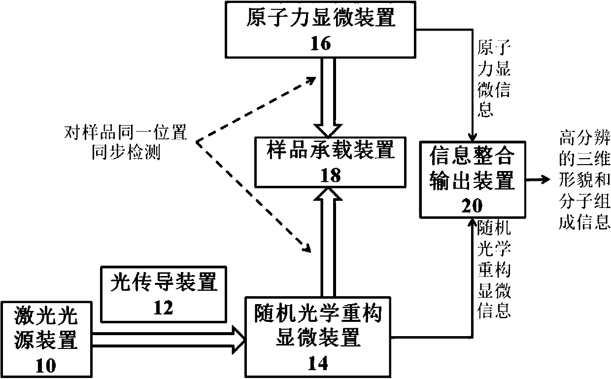
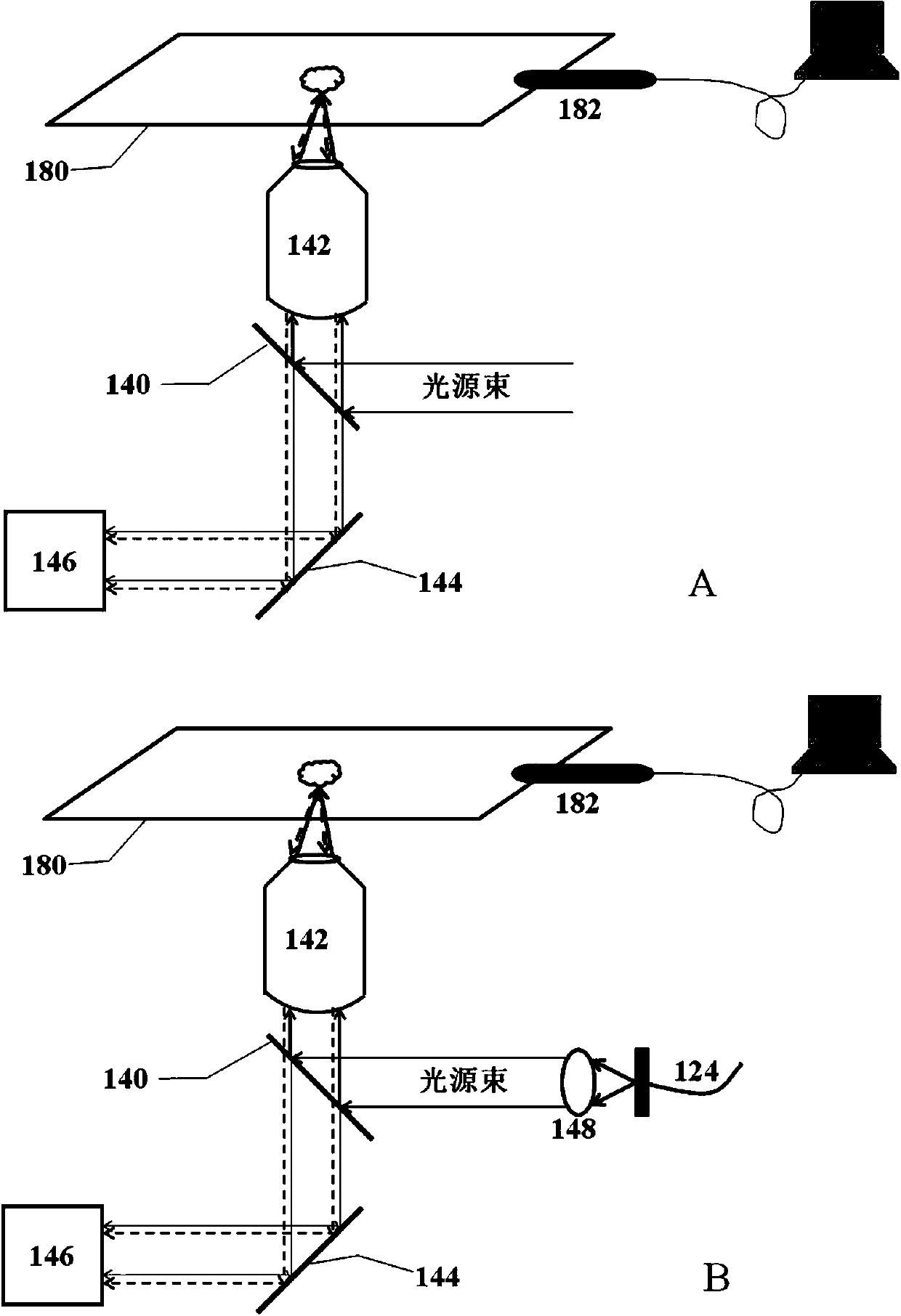
[0052] Conduct high-resolution morphology and composition studies on cell membranes, fix biofilm samples on the sample stage on the surface of the substrate, and perform antibody-labeled staining. The orientation controller in the sample carrying device controls the movement and rotation of the sample stage in three-dimensional directions, so as to select the position of the sample to be measured.
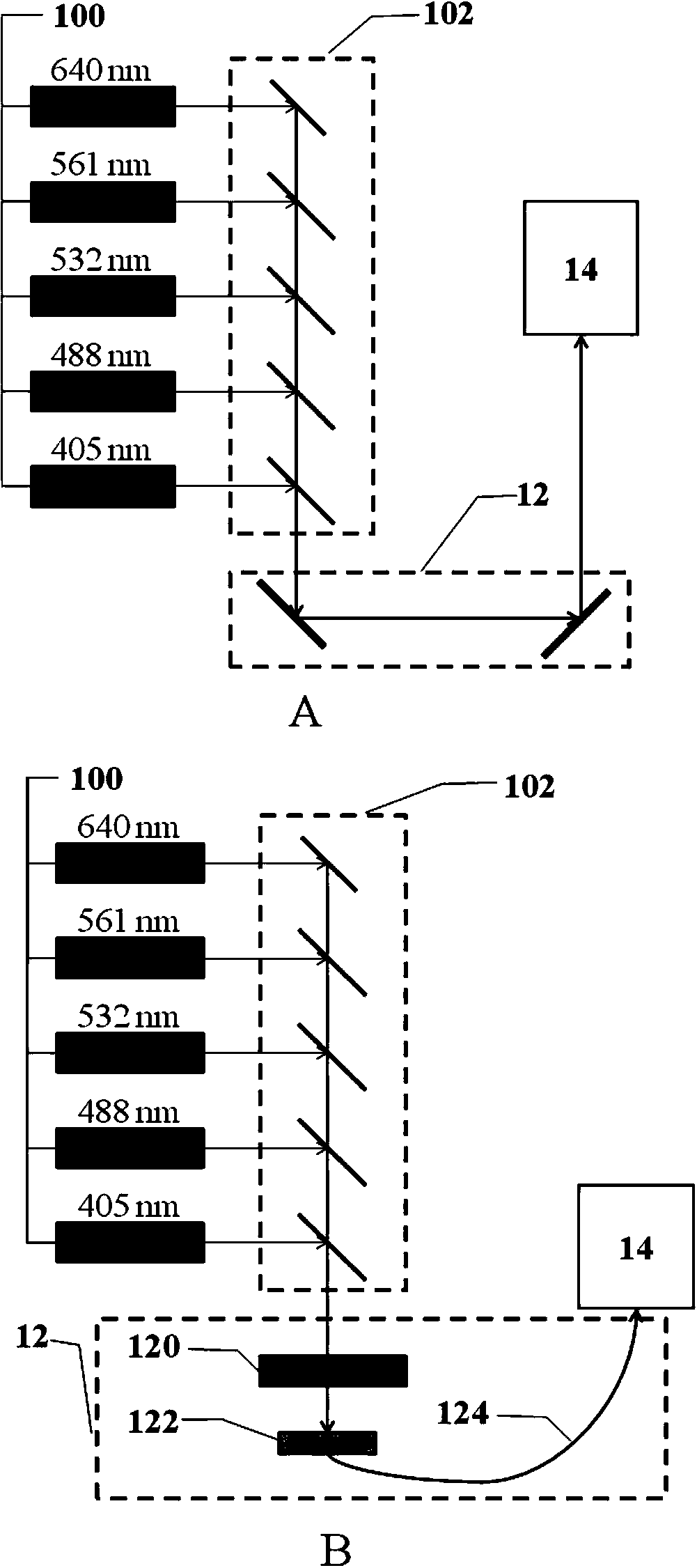
[0053] Five laser emitters respectively emit light with wavelengths of 640nm, 561nm, 532nm, 473nm and 405nm, which are converged into a light source beam by five dichromatic mirrors. The light source beam passes through the optical path attenuator and fiber optic coupler, and is guided by the optical fiber into the random reconstruction fluorescence microscope. The protein in the cell membrane sample is optically positioned by the random reconstruction fluorescence microscope, and the imaging result is output through the computer. Simultaneously with fluorescence imaging, the atomi...
PUM
 Abstract
Description
Claims
Application Information