Cervix uteri single cell image segmentation algorithm

An image segmentation and cervical cell technology, applied in the field of medical cell image processing, can solve the problems of inconsistent staining color, overlapping and adhering cells, containing impurities, etc., to improve the accuracy and efficiency of segmentation, and simplify the complex process.

Inactive Publication Date: 2015-10-21
GUANGXI NORMAL UNIV
View PDF3 Cites 14 Cited by
  • Summary
  • Abstract
  • Description
  • Claims
  • Application Information

AI Technical Summary

Problems solved by technology

[0004] The purpose of the present invention is to provide a cervical single-cell image segmentation algorithm for the deficiencies in the prior art. The cervical single-cell image segmentation algorithm of this adaptive threshold and ray gray gradient active contour model can avoid cell overlap in traditional segmentation methods Adhesion, inconsistent staining color and impurities in the background can improve the segmentation accuracy and efficiency of normal cervical single cells and cancerous cervical single cells

Method used


Image

  • Cervix uteri single cell image segmentation algorithm
  • Cervix uteri single cell image segmentation algorithm
  • Cervix uteri single cell image segmentation algorithm

Examples


Embodiment

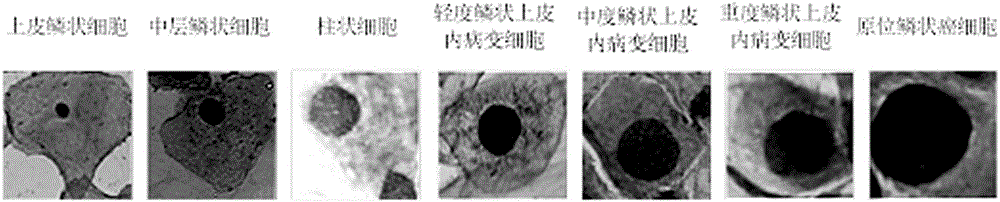
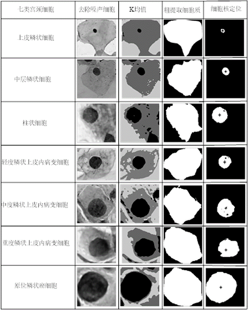
[0028] see figure 1 , derived from the Herlev cervical single-cell image dataset (http: / / labs.fme.aegean.gr / decision / downloads), the Herlev cervical single-cell image dataset was provided by the Technical University of Denmark and Herlev University Hospital ( Herlev University Hospital) jointly developed, the image resolution is 0.21 microns / pixel, a total of 917 cervical single cell images, the data set contains 7 types of cervical single cells, namely: normal columnar cells, normal middle cells, normal superficial cells, light Squamous intraepithelial lesion cells, moderate squamous intraepithelial lesion cells, severe squamous intraepithelial lesion cells, squamous cell carcinoma cells, 7 types of cervical single cell samples; this example randomly selects seven types of cervical single cell images authenticating.

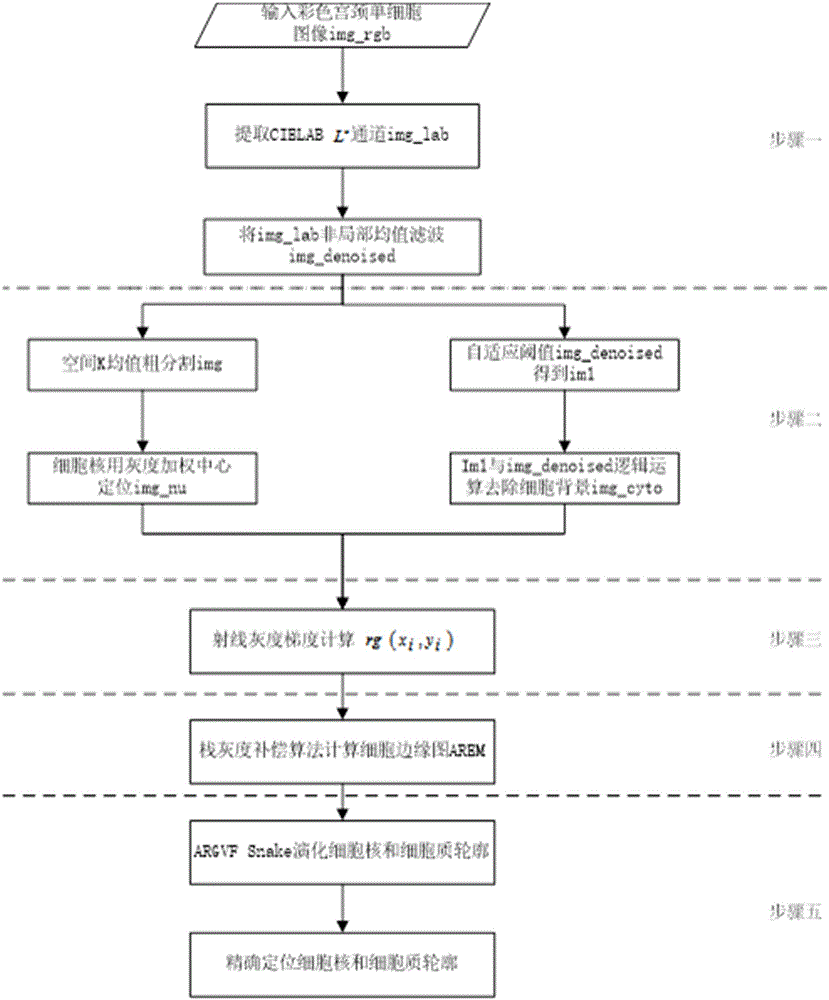
[0029] see figure 2 , the process includes a cell image preprocessing module, a rough segmentation module, a cell edge map module and a cell contour precise ...


PUM


Abstract

The invention discloses a method of a cervix uteri single cell image segmentation algorithm, and includes the steps of: 1) performing preprocessing on a cervix uteri single cell image to enhance a cell boundary; 2) removing a background of the cervix uteri cell image; 3) determining a gray scale gradient of a cell nucleus and cytoplasm along a ray direction; 4) using a stack gray scale difference compensation algorithm to optimize the ray gray scale gradient in the Step 3), and determining an edge map AREM of the cytoplasm and the cell nucleus of the cervix uteri single cell according to the noise-removed cervix uteri single cell image; and 5) applying gradient vector field GVF Snake model evolution to the determined edge map AREM of the cervix uteri single cell image to accurately locate profiles of the cytoplasm and the cell nucleus of the cervix uteri cell. The method simplifies a complex process of a traditional segmentation algorithm, solves the problems that in the traditional segmentation method cells overlap and adhere, dyed color is inconsistent and a background contains impurities, and improves the segmentation accuracy degree and segmentation efficiency of a normal cervix uteri single cell and a cancerous cervix uteri single cell.

Description

technical field [0001] The invention relates to the field of medical cell image processing, in particular to a cervical single-cell image segmentation algorithm based on an adaptive threshold value and a ray gray gradient active contour model. Background technique [0002] Cervical cancer has become one of the malignant tumors with the highest incidence rate, seriously endangering women's health. Because there are no obvious symptoms in the early stage of cervical cancer, the symptoms in the late stage are obvious but it is very difficult to cure. Therefore, early diagnosis of cervical cancer is the key to physician diagnosis and treatment. In modern medicine, cancer therapy through therapeutic cells is a new direction of development. In a type of computer-aided automatic diagnosis system represented by the cervical single-cell image processing system, the accurate segmentation of the nucleus and cytoplasm from the cell image is the basis for subsequent quantitative analys...

Claims


Application Information

Patent Timeline
Patent Type & Authority Applications(China)
IPC IPC(8): G06T7/00
CPCG06T7/0012G06T2207/30024
Inventor 刘艳红罗晓曙陈锦
Owner GUANGXI NORMAL UNIV
Why Eureka
Try Eureka