
Method for isolating and culturing endothelium from human adipose tissue for clone formation of cells

A technique for separating and culturing adipose tissue, which is applied in the field of cell biology to achieve the effects of short separation and culturing time, simple operation, and good angiogenesis

Active Publication Date: 2015-12-16
SICHUAN NEO LIFE STEM CELL BIOTECH
View PDF3 Cites 3 Cited by
  • Summary
  • Abstract
  • Description
  • Claims
  • Application Information

AI Technical Summary

Problems solved by technology

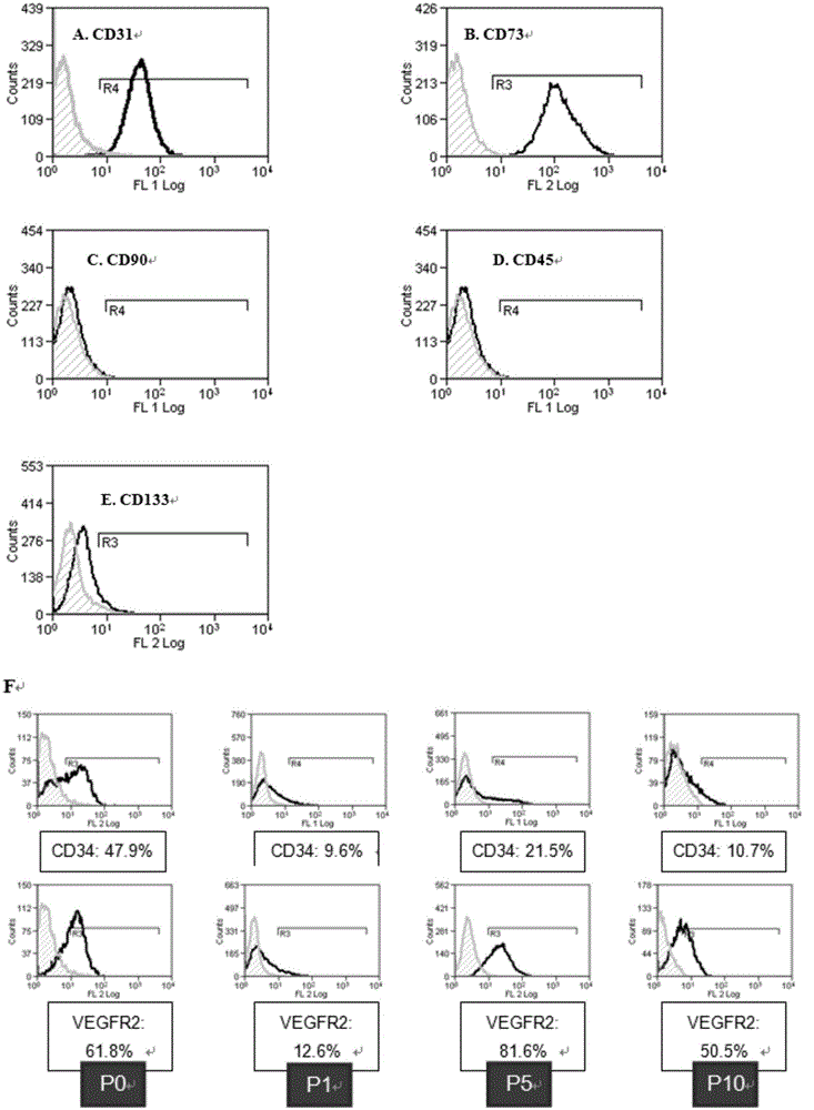
At present, there is no report on the isolation of ECFC from human adipose tissue. It is of great significance to isolate ECFC with strong proliferative ability from adipose tissue.

Method used


Image

  • Method for isolating and culturing endothelium from human adipose tissue for clone formation of cells
  • Method for isolating and culturing endothelium from human adipose tissue for clone formation of cells
  • Method for isolating and culturing endothelium from human adipose tissue for clone formation of cells

Examples


Embodiment 1

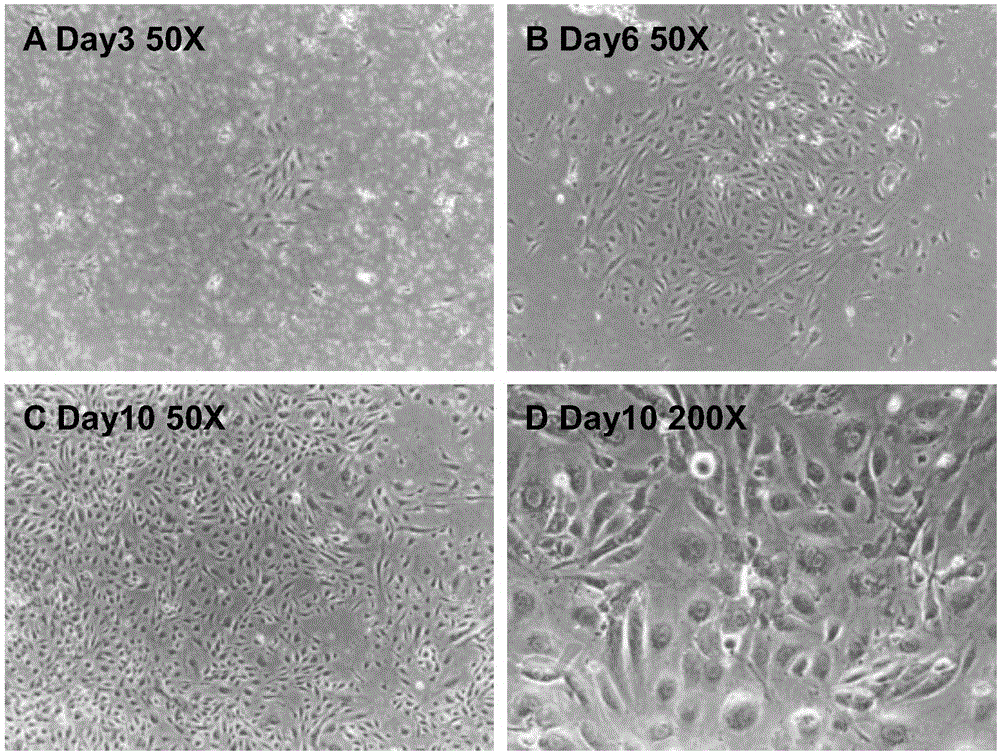
[0046] Example 1 Isolation and culture of endothelial clone-forming cells of the present invention

[0047] a. Take the adipose tissue, wash the adipose tissue with normal saline twice, remove the lower layer of liquid, and retain the upper layer of fat;

[0048] Add an equal volume of 0.2% type I collagenase solution, digest it in a shaker at 37°C for 1 hour, with an oscillation frequency of 150 rpm / min; put it in a centrifuge, centrifuge at 900 g for 5 minutes, discard the supernatant, and get Cell pellet

[0049] b. Add physiological saline to the cell pellet in step a, resuspend the cell cluster, centrifuge at 500g, and centrifuge for 5 minutes;

[0050] Discard the supernatant, resuspend the pellet in DMEM:F12 (1:1) containing 10% FBS, inoculate it in a T75 culture flask pre-coated with 0.1 mg / 1 mL type I rat tail collagen, and culture for 48 hours;

[0051] c. Replace the cell culture medium containing 10% FBS in step b with EGM2 medium, and culture for 1 day, and pavement-like c...

Embodiment 2

[0054] Example 2 Isolation and culture of endothelial clone-forming cells of the present invention

[0055] a. Take the adipose tissue, wash the adipose tissue with normal saline 3 times, remove the lower layer of liquid, and retain the upper layer of fat;

[0056] Add an equal volume of 0.1% type I collagenase solution, digest in a shaker at 37°C for 2 hours, with an oscillation frequency of 150 rpm / min; put it in a centrifuge, centrifuge at 500 g for 10 minutes, and discard the supernatant. Cell pellet

[0057] b. Add physiological saline to the cell pellet in step a, resuspend the cell cluster, centrifuge at 500g, and centrifuge for 5 minutes;

[0058] Discard the supernatant, resuspend the pellet in DMEM:F12 (1:1) containing 10% FBS, inoculate it in a T75 culture flask pre-coated with 0.1 mg / 1 mL type I rat tail collagen, and culture for 48 hours;

[0059] c. Replace the cell culture medium containing 10% FBS in step b with EGM2 medium, culture until the pavement-like cell colonies...

Embodiment 3

[0062] Example 3 Isolation and culture of endothelial clone-forming cells of the present invention

[0063] a. Take the adipose tissue, wash the adipose tissue with normal saline twice, remove the lower layer of liquid, and retain the upper layer of fat;

[0064] Add an equal volume of 0.5% type I collagenase solution, digest it in a shaker at 37°C for 0.5 hours, with an oscillation frequency of 150 rpm / min; put it in a centrifuge, centrifuge at 900g for 5 minutes, discard the supernatant, and get Cell pellet

[0065] Steps b, c, d are the same as in Example 2.


PUM


Abstract

The invention provides a method for isolating and culturing endothelium from human adipose tissue for clone formation of cells. The method comprises the steps of a, taking adipose tissue and performing washing, digestion and centrifuging to obtain cell sedimentation; b, cleaning the cell sedimentation in the step a, placing the cell sedimentation in a cell culture medium containing 10% FBS, and culturing the cell sedimentation for 48 h; c, replacing the cell culture medium containing 10% FBS in the step b with an EGM2 culture medium, performing culture till paving stone sample cell clone groups appear, and replacing the solution every 2-3 days in the culture period; d, replacing the EGM2 culture medium with a mixed culture medium every 2 days till the cell merging degree reaches 80-90%, and the cells are obtained. The invention further provides the prepared cells formed by clone formation. The isolation method is applied, endothelium can be efficiently obtained from the adipose tissue for clone formation to obtain the cells, and the method is easy to implement and short in isolation and culture time and has the good application prospect.

Description

Technical field [0001] The invention belongs to the field of cell biology, and specifically relates to a method for separating and culturing endothelial clone-forming cells from human adipose tissue. Background technique [0002] Endothelial Progenitor Cells (EPC), also known as angioblasts, are a type of precursor cells that can proliferate and differentiate into Endothelial Cells (EC). EPC participates in the process of angiogenesis and blood vessel formation in the body, and has broad application prospects in repairing myocardial infarction, skin damage, treating ischemic diseases, vascular tissue engineering and tumor treatment. [0003] EPC has different subtypes at different stages of cell differentiation. Generally, EPC is divided into early EPC (including CFU-EC, CAC) and late EPC (ie endothelial clone forming cells, abbreviated as ECFC). The literature provides different EPC subtypes. Identification method [1. JinHuretal, Characterization of TwoTypes of Endothelial Progen...

Claims


Application Information

Patent Timeline
IPC IPC(8): C12N5/074
Inventor 范兆心陈强赖真阳
Owner SICHUAN NEO LIFE STEM CELL BIOTECH
Learn More