Operative positioning system

A positioning system and surgical technology, applied in the field of surgical positioning systems, can solve problems such as volume limitations and difficulty in meeting configuration requirements, and achieve the effect of reducing requirements

Active Publication Date: 2016-01-06
BEIJING YIQIANCHUANG TECH CO LTD
View PDF5 Cites 3 Cited by
  • Summary
  • Abstract
  • Description
  • Claims
  • Application Information

AI Technical Summary

Problems solved by technology

[0008] CN201310298142 patent has other problems: 1 / The cloud server function of this technology is placed behind, and the function of the cloud server is placed in the last link of the process - the stage of postoperative evaluation
The above two points make a large amount of calculation work must be completed by the local processor, which puts forward certain requirements for the configuration of the local processor.
However, mobile devices and wearable devices are inherently limited by their size. Compared with desktop computers or even workstations, it is not easy to meet these configuration requirements.

Method used


Image

  • Operative positioning system

Examples


Embodiment 1

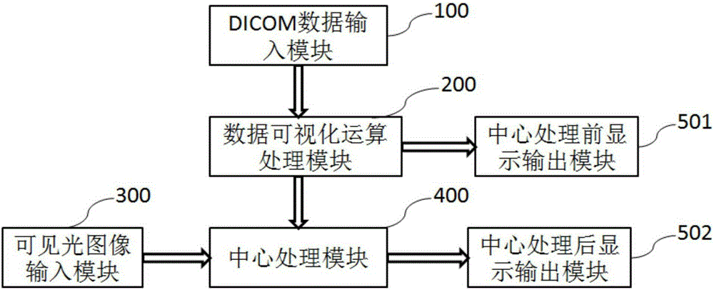
[0031] A non-enhanced CT scan of a patient with renal diverticulum found a stone buried in the renal parenchyma (diverticulolithiasis), and contrast-enhanced CT showed the inner lumen structure of the kidney and ureter (collecting system). The doctor uploads the CT data of the patient to the cloud server in the form of DICOM files. After data visualization processing, the 3D model data of the patient's kidney and the position data of the stone in the kidney are transmitted to the central processing module. The central processing module accepts the image data from the flexible ureteroscope camera and the 3D data from the data visualization processing module. Through registration and fusion, it can determine which relative position of the scene in the lens is in the patient's 3D model, and where it is buried. The required path of travel for stones in a diverticulum. In this way, along the path suggested by the central processing module, diverticular stones buried in the tissue ...

Embodiment 2

[0033]A patient with kidney tumors needs laparoscopic partial nephrectomy. The doctor uploads the patient's kidney CT data to the cloud server in the form of a DICOM file. After data visualization processing, the 3D model data of the patient's kidney, the location data of the tumor in the kidney and the location of the kidney blood vessels are transmitted to the central processing module. The central processing module is also located in the cloud, accepting the image data from the laparoscopic camera and the 3D data from the data visualization processing module. Through registration and fusion, it can determine the direction of blood vessels supplying kidney tumors under the kidney capsule, which can be displayed on the on wearable devices (glasses). The doctor thus selectively blocks only the blood vessels supplying the kidney tumor and completes the operation. It avoids the need to block larger arteries and veins in conventional methods, resulting in more extensive renal t...

Embodiment 3

[0035] A patient with peripheral lung cancer needs a bronchoscopic biopsy. The doctor uploads the patient's DICOM file to the cloud server. After data visualization processing, the 3D model data of the patient's lungs and bronchi at all levels, the location of the tumor in the lung and the location of blood vessels around the tumor are transmitted to the central processing module. The central processing module accepts the image data from the bronchoscope camera and the 3D data from the data visualization processing module. Through registration and fusion, it can determine which relative position of the bronchus lens is in the patient's 3D model, and which bronchi need to pass through to reach the tumor. Fork, whether there are blood vessels around the tumor that need to be avoided during biopsy, etc. It can even assist in the identification and selection of tumor margins for biopsy, because the tumor margins have a higher detection rate of cancer cells than the tumor center (...


PUM


Abstract

The invention discloses an operative positioning system. The operative positioning system completes positioning through direct comparison between real-time visible light images and non-real-time imageology images. The operative positioning system comprises a DICOM (digital imaging and communications in medicine) data input module, a data visualization operation processing module, a visible light image input module, a central processing module and an image display output module. The operative positioning system has the advantages that on the basis of imageology data, a preoperative 3D (three-dimensional) non-real-time image model is generated and then is fused with an intra-operative real-time camera image, so that requirements on intra-operative equipment can be lowered, for example, special laparoscopy ultrasonic equipment or endoscope ultrasonic equipment is unneeded, and only routine preoperative imaging results need to be used; according to the method, theoretically, a 100% lesion part can be displayed on the 3D model surely, and a camera only needs to focus on corresponding positions of operative instruments and symbolic anatomical structures on a 3D map.

Description

technical field [0001] The invention relates to the technical field of medical instruments, in particular to an operation positioning system. Background technique [0002] Today's medical examination and treatment has entered the era of minimally invasive. Laparoscopes, endoscopes (such as gastroscopes, colonoscopes, bronchoscopes) and surgical robots are all representatives of minimally invasive technologies. Among these representative technologies, various cameras are the main observation tools. They replace the human eye and are mainly used to perform two tasks: 1. Identify lesions and the location of lesions on the human body; 2. Identify surgical instruments and their locations on the human body. [0003] Surgical instruments are relatively large, and it is not difficult for the camera to identify them. However, it is difficult for the camera to identify lesions, especially early lesions. The reasons are: 1. The camera imaging technology uses visible light. Visible...

Claims


Application Information

Patent Timeline
Patent Type & Authority Applications(China)
IPC IPC(8): A61B19/00
Inventor 樊昊
Owner BEIJING YIQIANCHUANG TECH CO LTD
Why Eureka
Try Eureka