A corneal biomechanical detection instrument and its application method

A biomechanics and testing instrument technology, applied in the field of corneal biomechanical testing instruments, can solve the problems of inability to take continuous pictures of corneal images, no difference in intensity, inaccurate testing results, etc., and achieve improved corneal biomechanical properties and corneal elasticity The effect of reducing, ensuring accuracy

Active Publication Date: 2019-03-19
JINAN SANWEI MEDICAL INSTR CO LTD
View PDF8 Cites 0 Cited by
  • Summary
  • Abstract
  • Description
  • Claims
  • Application Information

AI Technical Summary

Problems solved by technology

However, in actual use, it is often necessary to test multiple times and the results of each test are significantly different, and because the biomechanical properties of the cornea can be improved after the cornea is cross-linked by ultraviolet rays, those who have undergone ultraviolet cross-linking before using the above two instruments for detection In the case of cornea, there is often a problem that there is no difference between the intensity after surgery and the intensity before surgery, or the data deteriorates
After analysis, it is believed that the traditional non-contact detection method cannot continuously take pictures when using imaging technology to obtain corneal images, that is to say, the collection of corneal deformation information is intermittent and discontinuous during detection, so the collected information exists deviation, and due to the existence of the deviation of the collected information, the final detection result will be inaccurate, which is the deficiency in the prior art

Method used


Image

  • A corneal biomechanical detection instrument and its application method
  • A corneal biomechanical detection instrument and its application method
  • A corneal biomechanical detection instrument and its application method

Examples


Embodiment Construction

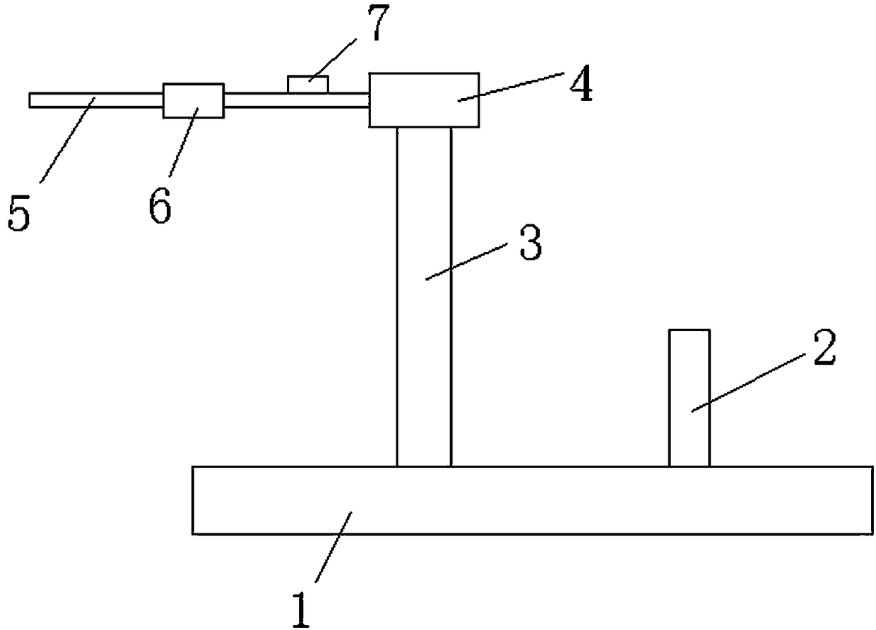
[0038] The technical scheme of the present invention will be described in detail below in conjunction with the accompanying drawings.

[0039] At present, the corneal elasticity of patients with keratoconus and corneal ectasia is reduced, which leads to the inability of the cornea to resist intraocular pressure, resulting in corneal deformation, changes in the refractive state of the eyeball, high myopia, and severe cases leading to corneal rupture and blindness. The existing non-invasive treatment of keratoconus and corneal ectasia is mainly corneal ultraviolet cross-linking, which can improve the biomechanical properties of the cornea, but so far there is no instrument that can effectively detect the improvement of corneal elasticity after ultraviolet cross-linking treatment .

[0040] The corneal biomechanics detection instrument and method provided by the present invention can detect the improvement of the corneal elasticity of the cornea treated by ultraviolet cross-linki...


PUM


Abstract

The invention belongs to the technical field of ophthalmological detection and particularly relates to an instrument for detecting cornea biomechanics and an application method of the instrument. The instrument comprises a power device. The power device is connected with a probe through a power transmission mechanism. The probe is used for detecting the biomechanical performance of corneas and provided with a pressure sensor and a displacement sensor. The instrument for detecting cornea biomechanics is capable of increasing detection data accuracy and simple and reliable in detection process.

Description

technical field [0001] The invention belongs to the technical field of ophthalmology detection, and in particular relates to a corneal biomechanical detection instrument and a use method thereof. Background technique [0002] The cornea is a complex biomechanical composite structure whose properties depend on structural subunits and patterns of assembly. In the cornea, only the Bowman's membrane and the stroma are collagen tissue layers, which are the main structural components that provide corneal strength; the corneal epithelium contributes little to the corneal strength, and the curvature of the cornea after corneal refractive surgery removes the corneal epithelium is almost No effect; the stretchability and low strength of the Descemet's membrane make it stretchable under a wide range of intraocular pressure, and it can well buffer and protect the corneal stroma that may cause damage to the corneal endothelium damage. [0003] The structural material and mechanical pro...

Claims


Application Information

Patent Timeline
Patent Type & Authority Patents(China)
IPC IPC(8): A61B3/107
CPCA61B3/107
Inventor 李志伟牟国营张仝
Owner JINAN SANWEI MEDICAL INSTR CO LTD
Why Eureka
Try Eureka