Biological tissue blood flow, blood oxygen and blood volume multi-parameter detection method and device

A technology of biological tissue and detection method, applied in the field of biomedical imaging, can solve the problem of high cost

Active Publication Date: 2016-03-16
HUAZHONG UNIV OF SCI & TECH
View PDF5 Cites 36 Cited by
  • Summary
  • Abstract
  • Description
  • Claims
  • Application Information

AI Technical Summary

Problems solved by technology

[0006] This system uses a beam splitter and an optical filter to split the reflected light carrying the signal according to different wavelengths, and then collects it with multiple monochrome cameras. This method can avoid time-sharing

Method used


Image

  • Biological tissue blood flow, blood oxygen and blood volume multi-parameter detection method and device
  • Biological tissue blood flow, blood oxygen and blood volume multi-parameter detection method and device
  • Biological tissue blood flow, blood oxygen and blood volume multi-parameter detection method and device

Examples


Embodiment Construction

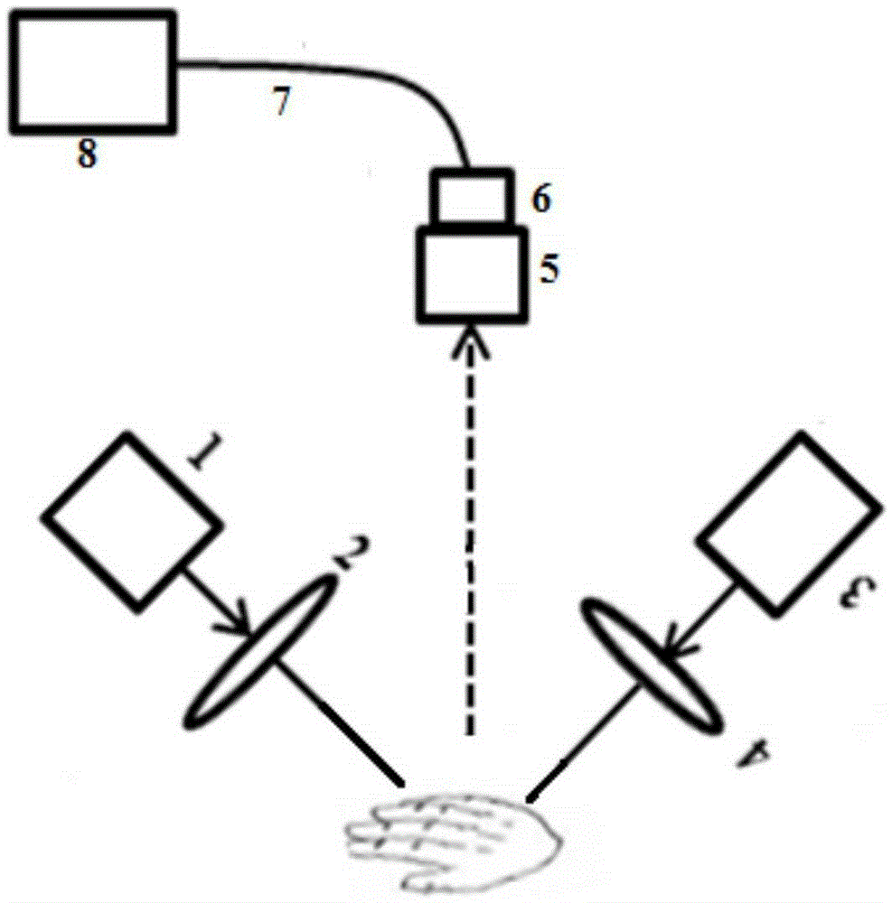
[0045] Such as figure 1 Shown: the device includes a first laser 1, a first beam expander 2, a second laser 3, a second beam expander 4, an imaging lens 5, a color area array CCD or color area array CMOS camera 6, a data transmission line 7 and computer8.

[0046] The first laser 1 (with an optical power of 20 milliwatts and a wavelength of 635 ± 5 nanometers) and the second laser (with an optical power of 20 milliwatts and a wavelength of 530 ± 5 nanometers) 3 pass through the first beam expander 2 and the second laser respectively. The second beam expander 2 obtains the divergent laser beam and irradiates it on the biological tissue sample at the same time, such as figure 1 The back of the human hand is shown. The imaging lens 5 images the irradiated biological tissue onto the color camera 6, and the data transmission line 7 (gigabit network cable) continuously captures the images captured by the color camera 6 and saves them to the computer 8 for data processing.

[0047...


PUM


Abstract

The invention discloses a biological tissue blood flow, blood oxygen and blood volume multi-parameter detection method and device. According to the method, two laser beams, differing in wavelength, simultaneously irradiate a biological tissue; and multiple frames of biological tissue colored images, which are illuminated by the laser beams, are continuously acquired by a photoelectric imaging system. The light intensity values of a red channel and a green channel on each pixel of the images are obtained for respectively constituting a red channel image and a green channel image. By virtue of a laser speckle blood flow imaging data analysis method and a dual-wavelength spectroscopic data analysis method, the red channel image and the green channel image are processed, so that two-dimensional spatial distribution and dynamic variation information thereof of a plurality of physiological parameters including blood flow, oxyhemoglobin concentration and reduced hemoglobin concentration of the corresponding biological tissue are obtained. The method and the device disclosed by the invention solve the shortcoming that a multi-wavelength time-sharing imaging based multi-parameter blood flow imaging system has difficulty in achieving the synchronization of accurate time and also avoid the shortcoming that a multi-camera shooting based multi-parameter blood flow imaging system is high in cost due to the use of a plurality of cameras.

Description

technical field [0001] The patent of the present invention belongs to the field of biomedical imaging, and specifically relates to an easy-to-use and low-cost multi-parameter detection method and device, which can realize real-time imaging of multiple hemodynamic information of biological tissues such as human skin and cerebral cortex, and Continuous monitoring. Background technique [0002] The multi-parameter monitoring of biological tissue hemodynamics is very important for studying the physiological state of living organisms. The general multi-parameter blood flow imaging system is realized by combining basic optical imaging methods such as various fluorescence imaging, endogenous signal optical imaging and laser speckle blood flow imaging, and these optical imaging are achieved by using incoherent or coherent The light source irradiates biological tissue, collects its reflected light, and analyzes the reflected light information to obtain hemodynamic parameters. Howev...

Claims


Application Information

Patent Timeline
IPC IPC(8): A61B5/026A61B5/1455
Inventor 李鹏程骆清铭王佳陆锦玲
Owner HUAZHONG UNIV OF SCI & TECH
Why Eureka
Try Eureka