Three-dimensional CT image segmentation method and three-dimensional CT image segmentation device

A CT image, two-dimensional image technology, applied in the field of three-dimensional CT image segmentation, can solve the problems of containing more and affecting the diagnostic accuracy, and achieve the effect of improving the diagnostic accuracy and accuracy.

Active Publication Date: 2016-04-20
SHANGHAI UNITED IMAGING HEALTHCARE
View PDF4 Cites 23 Cited by
  • Summary
  • Abstract
  • Description
  • Claims
  • Application Information

AI Technical Summary

Problems solved by technology

In the prior art, after the target parenchymal area is obtained, and in the process of finding out the lesion area in the target parenchymal area, since the target parenchymal area may contain other body tissues, resulting in Mistaking other body tissue parts as diseased areas, such as mistaking the spinal column area containing the human body in the lung parenchyma area as a pulmonary nodule area, will eventually lead to more false positive lesion areas in the lesion diagnosis results, affecting the accuracy of diagnosis

Method used


Image

  • Three-dimensional CT image segmentation method and three-dimensional CT image segmentation device
  • Three-dimensional CT image segmentation method and three-dimensional CT image segmentation device
  • Three-dimensional CT image segmentation method and three-dimensional CT image segmentation device

Examples


Embodiment Construction

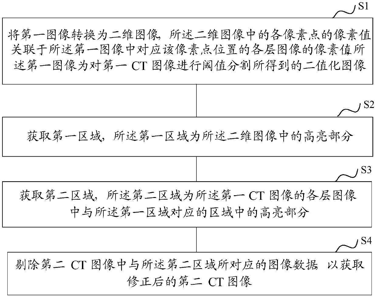
[0037] In the prior art, there is a problem that the lesion diagnosis result of human tissue obtained by using the image segmentation method contains many false positive lesion areas.

[0038] In order to solve the above problems, the technical solution of the present invention provides a method for segmenting three-dimensional CT images.

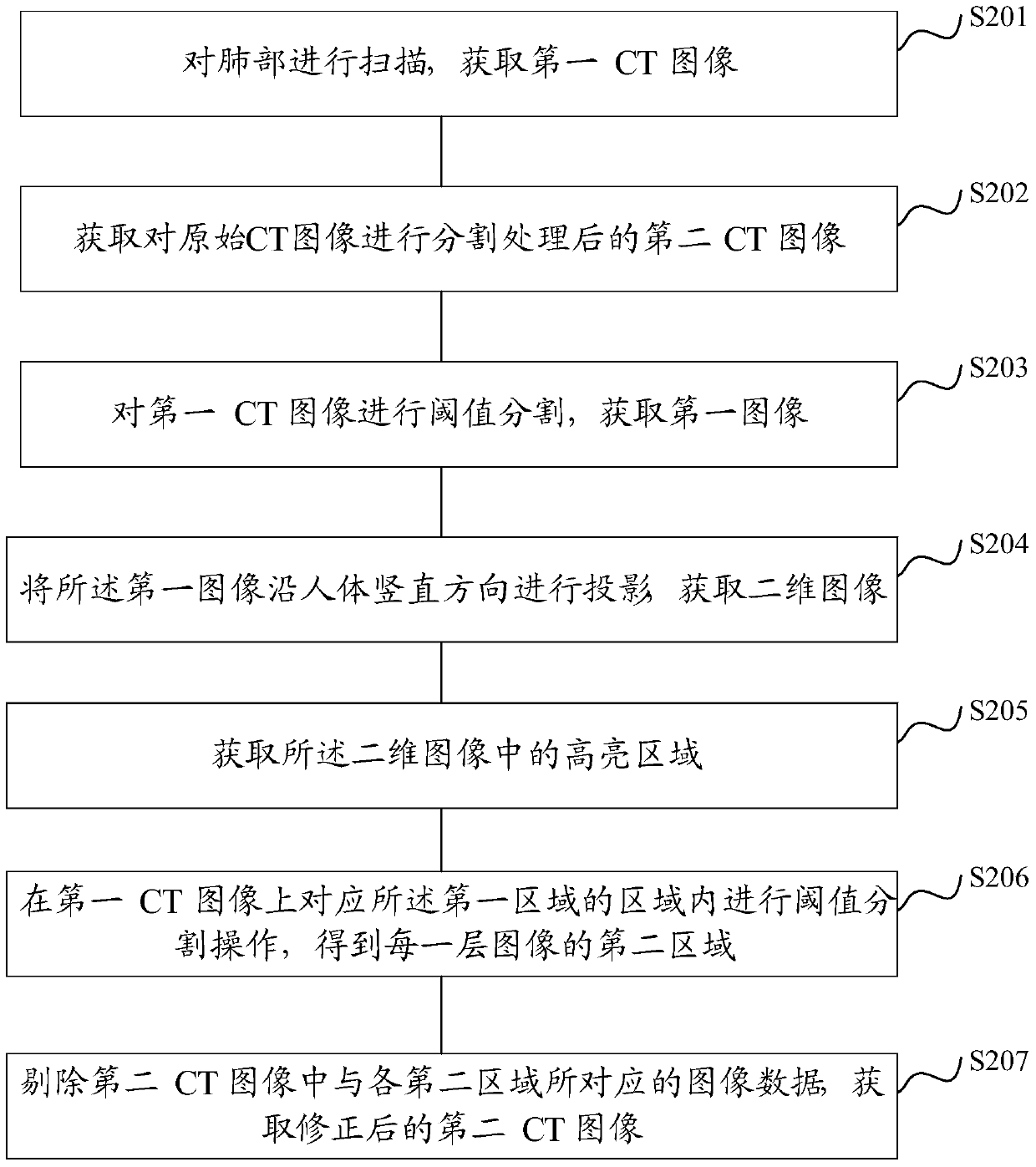
[0039] figure 1 It is a schematic flowchart of the segmentation method of the three-dimensional CT image provided by the technical solution of the present invention. First, step S1 is performed to convert the first image into a two-dimensional image, and the pixel value of each pixel in the two-dimensional image is associated with the pixel value of each layer image corresponding to the pixel position in the first image.

[0040] The first image is a binarized image of the region of interest obtained by performing threshold segmentation on the first CT image, and the first CT image is an original CT image taken according to a diagnosis req...


PUM


Abstract

The invention discloses a three-dimensional CT image segmentation method and a three-dimensional CT image segmentation device. The three-dimensional CT image segmentation method is characterized in that a first image can be converted into a two-dimensional image, and the first image is a binary image acquired by adopting threshold segmentation of a first CT image; a first area is acquired, and is the high light part of the two-dimensional image; a second area is acquired, and is the high light parts of the areas of the images of different layers of the first CT image corresponding to the first area; image data of a second CT image corresponding to the image data of the second area can be eliminated, and the corrected second CT image can be acquired. The method provided by the invention is advantageous in that the bone and other normal-appearing areas of the second CT image can be effectively eliminated, and the accuracy of the human tissue segmentation result can be improved, and therefore the accuracy of the disease diagnosis result including the diseased areas can be guaranteed, and the diagnosis accuracy can be improved.

Description

technical field [0001] The invention relates to the technical field of medical image processing, in particular to a method and device for segmenting three-dimensional CT images. Background technique [0002] Computed tomography (CT, Computed Tomography) is widely used in the diagnosis of diseases and has become one of the important means for doctors to conduct pathological analysis, anatomical research and functional evaluation. An important area of ​​study for efficiency and proficiency. [0003] In the process of disease diagnosis, it is usually necessary to segment the region of interest of the doctor from the three-dimensional CT image, and the region of interest (Region of Interest, ROI) usually refers to the target tissue, such as the solid region or focus of the chest, lung, etc. Regions, accurate segmentation of regions of interest in CT scan images, identification and classification of target tissues, and diagnosis of diseases are all crucial. [0004] Image segme...

Claims


Application Information

Patent Timeline
Patent Type & Authority Applications(China)
IPC IPC(8): G06T7/00A61B6/03
Inventor 王季勇
Owner SHANGHAI UNITED IMAGING HEALTHCARE
Why Eureka
Try Eureka