Primary isolated culture method for dairy cow mammary epithelial cells

A technique for separating and culturing mammary gland epithelial cells, which is applied in the field of rapid separation of dairy cow mammary gland epithelial cells, can solve the problems of no glandular epithelium in shape, and other problems that have not been obtained, and achieve the effect of shortening the time for migration

Inactive Publication Date: 2016-05-04
NORTHWEST A & F UNIV
View PDF4 Cites 12 Cited by
  • Summary
  • Abstract
  • Description
  • Claims
  • Application Information

AI Technical Summary

Problems solved by technology

Although these immortalized cell lines maintain part of the biological characteristics and functions similar to those of the original cells, they have morphological differentiation or no typical characteristics of glandular epithelium during use, and some have not yet been verified by other laboratories. testing, and only for exchange between laboratories, so far there is no commercial dairy mammary epithelial cell line available for researchers

Method used


Image

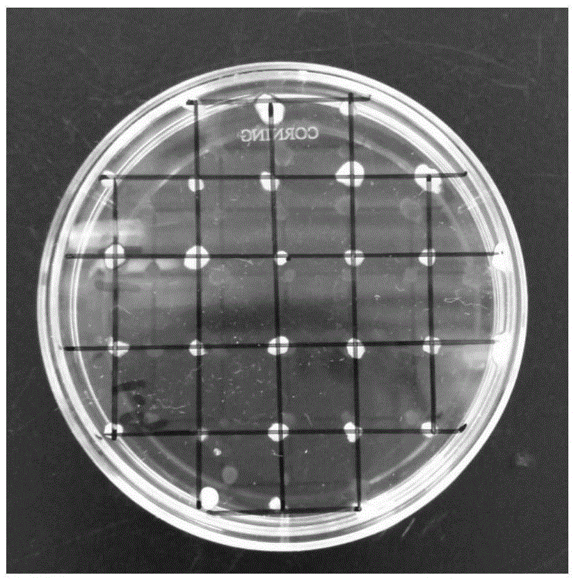
  • Primary isolated culture method for dairy cow mammary epithelial cells
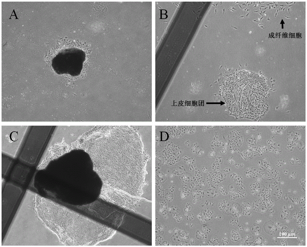
  • Primary isolated culture method for dairy cow mammary epithelial cells
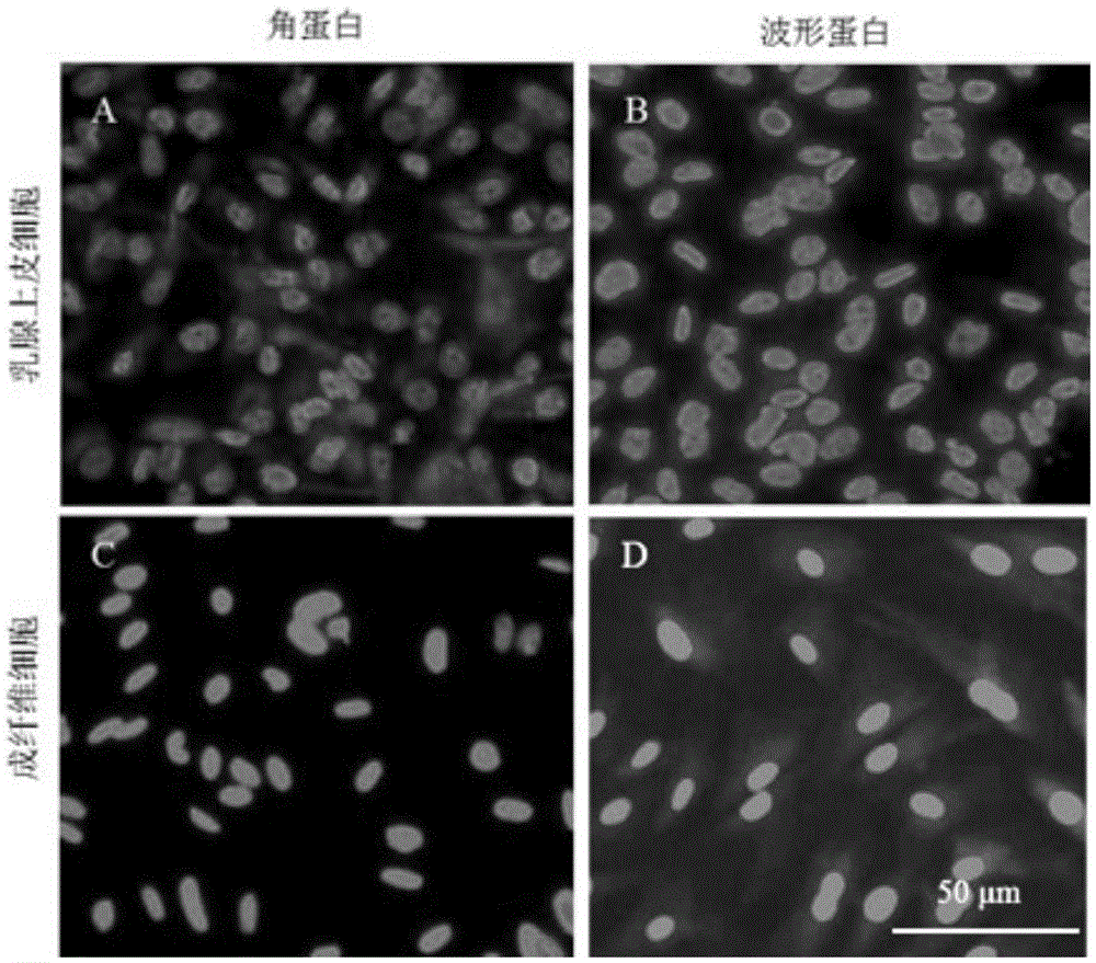
  • Primary isolated culture method for dairy cow mammary epithelial cells

Examples


Embodiment Construction

[0028] In order to make the purpose and technical solution of the present invention more complete and clear, the present invention will be described in detail below in conjunction with the accompanying drawings and embodiments. It should be understood that the embodiments described here are only used to explain the present invention, but not to limit the present invention.

[0029] (1.1) Preparation of tissue digestion solution (collagenase / hyaluronidase digestion solution)

[0030] Add 1mg collagenase (0.25~1.0FALGPAUnits / mg), 250μg hyaluronidase (400~1000Unit / mg), 100IU penicillin, 100μg streptomycin and 0.25μg amphotericin B to each milliliter of DMEM / F12 culture medium Prepared, sterilized by filtration and stored at -20°C, thawed before use and preheated to 37°C in a water bath.

[0031] (1.2) Cell culture medium

[0032] DMEM / F12 culture medium containing 10% fetal bovine serum by volume, 100 IU / mL penicillin, 100 μg / mL streptomycin and 0.25 μg / mL amphotericin B. Spec...


PUM


Abstract

The invention discloses a primary isolated culture method for dairy cow mammary epithelial cells. The primary isolated culture method comprises steps as follows: mammary tissue of a dairy cow in the lactation period is shorn into tissue blocks, the tissue blocks are digested with collagenase / hyaluronidase digestive juice, a culture dish is inoculated with the digested chylous tissue blocks for culture, cells get free from tissue on peripheries of the tissue blocks after 1-2 days and grow along the wall of the culture dish, fusiform fibroblast is removed with a mechanical scraping method every day during culture, and initial passage is performed when a large quantity of cobblestone-like mammary epithelial cells grow in clusters and in patches and are mutually fused after about 4-5 days. During initial passage, a little fibroblast which is probably mixed is eliminated with trysin with a differential digestive method, and the dairy cow mammary epithelial cells with high purity are obtained. The mammary epithelial cells can emigrate from chylous tissue in a short time; a little mixed fibroblast is removed mainly with a mechanical scraping method, and accordingly, the obtained mammary epithelial cells are seldom damaged and are high in purity.

Description

technical field [0001] The invention relates to the field of cell biology, in particular to a method for rapidly separating cow mammary gland epithelial cells. Background technique [0002] Cow mastitis is one of the most harmful and common diseases in dairy farming. The harm caused by dairy cow mastitis has led scholars at home and abroad to conduct in-depth research on mastitis from multiple perspectives and aspects such as etiology, pathogenesis, diagnosis, treatment and prevention, and the most commonly used research on mastitis is to establish A mastitis cell model of mammary epithelial infection in vitro. In addition, mammary gland epithelial cells are recognized as the target cells for making mammary gland bioreactors, that is, using mammary gland epithelial cells cultured in vitro, based on the transgenic technology platform, foreign genes are introduced into the animal genome and expressed in the animal mammary gland. Some valuable products, such as small molecule...

Claims


Application Information

Patent Timeline
Patent Type & Authority Applications(China)
IPC IPC(8): C12N5/071
CPCC12N5/0631C12N2509/00
Inventor 高明清张涌张文耀和桂良陈青徐彤
Owner NORTHWEST A & F UNIV
Why Eureka
Try Eureka