Monoclonal antibody with function of blocking human programmed death factors 1 (PD-1), encoding gene of monoclonal antibody and application thereof

A monoclonal antibody, PD-1 technology, applied in application, antibody, genetic engineering and other directions, to achieve the effect of high affinity and broad application prospects

Active Publication Date: 2016-05-11
大庆东竺明生物技术有限公司
View PDF2 Cites 32 Cited by
  • Summary
  • Abstract
  • Description
  • Claims
  • Application Information

AI Technical Summary

Problems solved by technology

Experiments confirm that blocking PD-1 signaling may be appli

Method used


Image

  • Monoclonal antibody with function of blocking human programmed death factors 1 (PD-1), encoding gene of monoclonal antibody and application thereof
  • Monoclonal antibody with function of blocking human programmed death factors 1 (PD-1), encoding gene of monoclonal antibody and application thereof
  • Monoclonal antibody with function of blocking human programmed death factors 1 (PD-1), encoding gene of monoclonal antibody and application thereof

Examples


Embodiment 1

[0025] Stable membrane expression of hPD-1

[0026]In order to obtain a cell line expressing hPD-1 protein on the cell membrane surface as an immunogen. Purchase the cDNA clone encoding the full-length open reading frame of human PD-1 (Beijing Yiqiao Shenzhou Biotechnology Co., Ltd. HG10377-CF, Genbank accession number NM-005018.2), and insert it into pCDNA3.1(+) (invitrogen) In the vector, after sequencing confirms that the coding frame of the hPD-1 gene is correct. The plasmid was electrotransformed into CHO-DG44 cells, pressurized screening, and cloned to obtain CHO-DG44 cells stably expressing hPD-1. Named hPD-1 / DG44.

Embodiment 2

[0028] animal immunity

[0029] Ten A / J mice aged 6-8 weeks were selected and immunized five times with an interval of 14 days between each immunization. 107 hPD-1 / DG44 cells were immunized each time, subcutaneously and intraperitoneally at multiple points. For the first immunization, an equal volume of Freund's complete adjuvant was mixed with cell suspension, and for the remaining 4 immunizations, Freund's incomplete adjuvant was mixed with cell suspension.

[0030] Configuration of cell culture medium

[0031] MD6 serum-free medium was used for culturing sp2 / 0 cells. After fusion, the cells are cultured in HAT medium, and the specific components are as follows: 10% fetal bovine serum and HAT are added to MD6 serum-free medium.

[0032] cell fusion

[0033] Three days after the final immunization, 6 mice with high titers were selected for cell fusion. Aseptically remove the mouse spleen and the pre-prepared sp2 / 0 cells, count the cells, mix them at a ratio of 10:1, and ...

example 3

[0041] Functional identification of anti-hPD1 monoclonal antibody

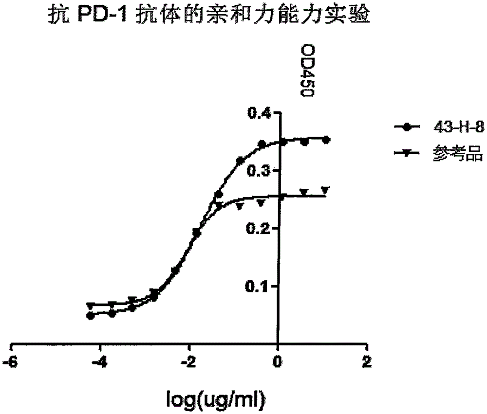
[0042] Affinity identification

[0043] A 96-well ELISA plate was coated with hPD-L1-Fc (RD); the blocking antibody was diluted 3 times as the primary antibody, and a total of 12 gradients were added to the hPD-L1-Fc-coated 96-well ELISA plate. Add goat anti-mouse IgG-Fc-HRP (SANTCruzBIotechnology) as the secondary antibody, add the chromogenic solution, and read the OD450 value after termination. EC50 concentrations were generated using Graphpad software. The EC50 of this antibody is 1.0665nM, and the EC50 of the positive control is 0.5344nM. It can be seen from the results that the screened antibody has obvious affinity for PD-1, and it is in the same order of magnitude as the positive control. (See figure 1 ).

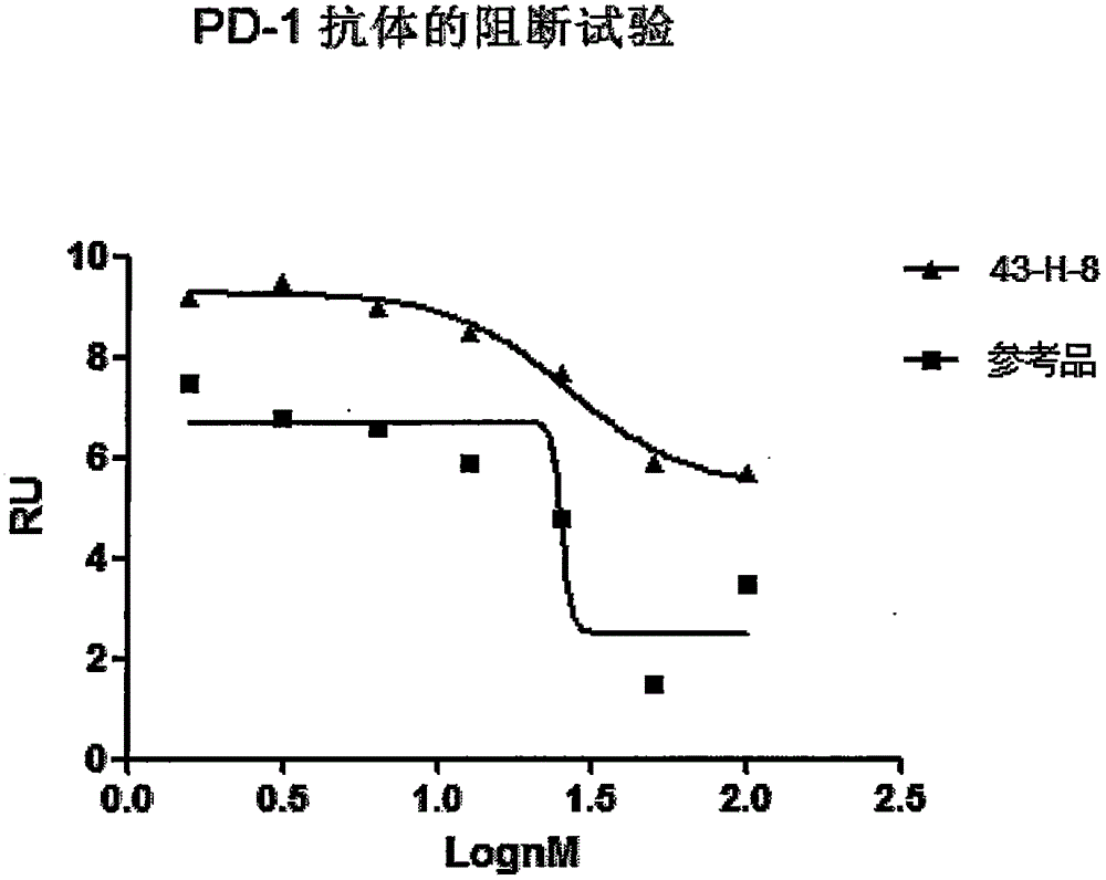
[0044] Functional Characterization of Antibody Blocking the Binding of hPD-1 and hPD-L1

[0045] A 96-well ELISA plate was coated with hPD-L1-Fc (RD); the purified hPD-1 antibody was diluted 4 ...


PUM


Abstract

The invention discloses a monoclonal antibody with a function of blocking human programmed death factors 1 (PD-1), an encoding gene of the monoclonal antibody and application thereof. The monoclonal antibody comprises heavy chains and light chains. Amino acid sequences of the heavy chains are shown as 20 bits to 132 bits of SEQ ID NO.3, and amino acid sequences of the light chains are shown as 20 bits to 130 bits of SEQ ID NO.4. The monoclonal antibody 43-H-8 with the function of blocking the human programmed death factors PD-1, the encoding gene of the monoclonal antibody and the application have the advantages that the monoclonal antibody 43-H-8 can be specifically bound with human PD-1 antigens, the medium effective concentration EC50 (50% effective concentration) is 1.0665nM, accordingly, PD-1/PD-L suppression signals can be specifically blocked, the monoclonal antibody 43-H-8 can be used as a blocker for PD-1 passages, and a novel medicine can be provided for tumor immunotherapy and treatment on chronic virus infectious diseases and autoimmune diseases.

Description

technical field [0001] The present invention relates to the technical field of biomedicine, in particular to the preparation, variable region sequence and application of anti-human PD-1 monoclonal antibody. Background technique [0002] Programmed cell death 1 (PD-1) is an immunosuppressive receptor expressed on the surface of activated T cells and B cells (Yao, Zhu et al. Advances in targeting cell surface signaling molecules for immune modulation. Nat Rev Drug Discov, 2013, 12(2): 130-146). When initially discovered, PD-1 was recognized as a member of the CD28 superfamily because of its structural similarity to the CD28 molecule. However, the PD-1 protein lacks the MYPPPY sequence that mediates the combination of CD28 / CTLA-4 and B7.1 / B7.2, and the FDPPPF sequence that mediates the combination of ICOS and ICOS-L, so it is structurally related to CD28, CTLA There are significant differences between -4 and ICOS. Therefore, the binding of PD-1 to the ligand is very specific ...

Claims


Application Information

Patent Timeline
IPC IPC(8): C07K16/30C12N15/13C12N15/79C12N5/10A61K39/395A61P35/00A61P31/12A61P37/02
Inventor 苏庆杨晓明刘云成杨岚哈卓王桂芬汤炜
Owner 大庆东竺明生物技术有限公司
Why Eureka
Try Eureka