Method for constructing cartilage tissues by aid of human urine cells

A cartilage tissue and urine cell technology, applied in the biological field, can solve the problems of limiting the application of bone marrow mesenchymal stem cells, pain at the sampling point, and large trauma.

Inactive Publication Date: 2016-06-01
GUANGZHOU INST OF BIOMEDICINE & HEALTH CHINESE ACAD OF SCI
View PDF3 Cites 11 Cited by
  • Summary
  • Abstract
  • Description
  • Claims
  • Application Information

AI Technical Summary

Problems solved by technology

In addition, the trauma to the body is greater when the material is collected, and there is pain at the sampling point, which also limits the clinical application of bone marrow mesenchymal stem cells

Method used


Image

  • Method for constructing cartilage tissues by aid of human urine cells
  • Method for constructing cartilage tissues by aid of human urine cells
  • Method for constructing cartilage tissues by aid of human urine cells

Examples


Embodiment 1

[0063] [Example 1] A method for reprogramming urine cells into induced pluripotent stem cells

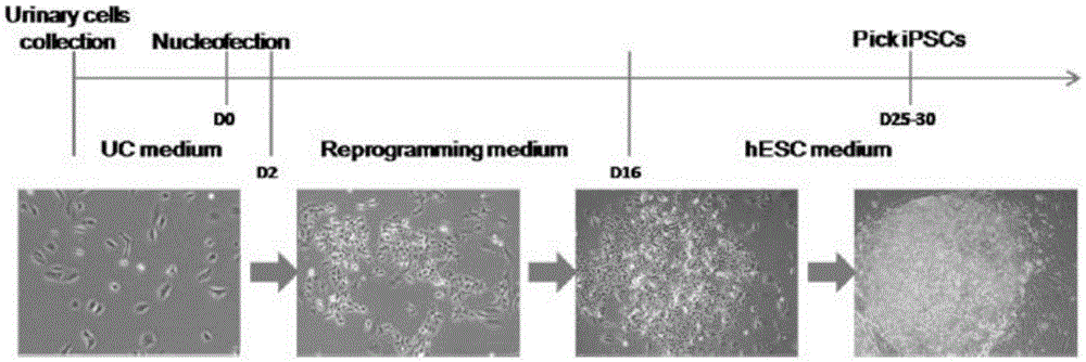
[0064] This embodiment relates to a method for generating iPS cells mediated by non-integrated attachment vectors derived from urine cells, which specifically includes the following steps ( figure 1 ):

[0065] 1) Introduce non-integrated episomal vectors (pEP4EO2SEN2K: 3.0 μg, pEP4EO2SET2K: 3.2 μg, pCEP4-M2L: 2.4 μg) expressing transcriptional regulators OCT4, SOX2, NANOG, KLF4 and LIN28 into 1 million urine cells, and then The cells were divided into three 10cm dishes pre-coated with matrigel and cultured with fibroblast medium;

[0066] 2) On the second day after transfection, the fibroblast medium was replaced with N2B27 conditioned medium (containing MEK inhibitor PD0325901, GSK3b inhibitor CHIR99021, TGF-b / Activin / Nodalreceptor inhibitor A-83-01, ROCK inhibitor HA- 100and human leukemia inhibitory factor (LIF), replace fresh medium every 2 days;

[0067] 3) On the 13th day ...

Embodiment 2

[0070] [Example 2] Optimizing the method for directed differentiation of iPS cells to mesenchymal stem cells

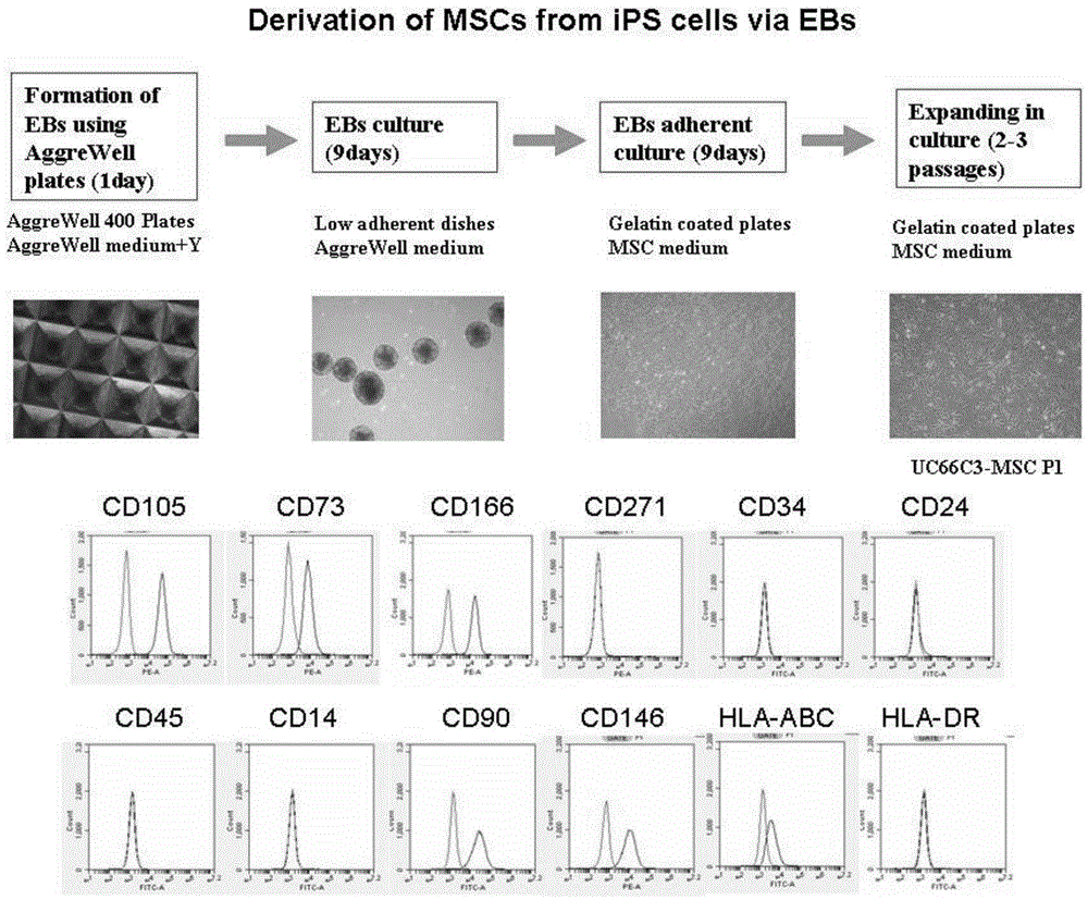
[0071] We optimized the method of directed differentiation of iPS cells to mesenchymal stem cells ( figure 2 ) includes: (1) iPS cells form embryoid bodies (EBs); (2) embryoid body suspension culture differentiates into mesoderm cells; (3) embryoid body adherent culture differentiated into mesoderm cells differentiates into mesenchymal stem cells .

[0072] We prepared embryoid bodies from iPS cells using AggreWell plates, which have microwells that can be used to aggregate iPS cells into embryoid bodies. When preparing embryoid bodies, add the single-cell suspension to the AggreWell culture plate, and then distribute the cells evenly in the microwells by centrifugation. After at least 24 hours of culture, the cells in each microwell are aggregated into clusters , prepared into embryoid bodies. The embryoid bodies obtained from AggreWell culture plates are very co...

Embodiment 3

[0073] [Example 3] A method of using urine cells to transdifferentiate into mesenchymal stem cells

[0074] This embodiment relates to a method for generating mesenchymal stem cells through transcription factor-mediated transdifferentiation of urine cells, which specifically includes the following steps ( Figure 5 ):

[0075] 1) Introduce non-integrated episomal vectors (pEP4EO2SEN2K: 3.0 μg, pEP4EO2SET2K: 3.2 μg, pCEP4-M2L: 2.4 μg) expressing transcriptional regulators OCT4, SOX2, NANOG, KLF4 and LIN28 into 1 million urine cells, and then The cells were divided into three 10cm dishes pre-coated with matrigel and cultured with fibroblast medium;

[0076] 2) On the second day after transfection, the fibroblast medium was replaced with N2B27 conditioned medium (containing MEK inhibitor PD0325901, GSK3b inhibitor CHIR99021, TGF-b / Activin / Nodalreceptor inhibitor A-83-01, ROCK inhibitor HA- 100and human leukemia inhibitory factor (LIF), replace fresh medium every 2 days;

[007...


PUM


Abstract

The invention provides a method for preparing cartilage tissues from artificial in-vitro sources by the aid of human urine cells in an in-vitro manner. The method includes steps of (1), a), reprogramming the human urine cells to generate induced pluripotent stem cells and directionally differentiating the induced pluripotent stem cells to obtain mesenchymal stem cells, or b), carrying out transdifferentiation to generate the mesenchymal stem cells; (2), cultivating the mesenchymal stem cells obtained at the step (1) in cartilage induced differentiation media to obtain the cartilage tissues from the artificial in-vitro sources. The method has the advantages that the cartilage tissues are constructed in the in-vitro manner and can be used for replacing damaged or lesion tissue cells, so that the purpose of repairing cartilage can be achieved, and the effective method is provided for constructing the cartilage tissues in the in-vitro manner and has huge application value.

Description

technical field [0001] The technology relates to the field of biotechnology, in particular to a method for constructing cartilage tissue by using human urine cells. Background technique [0002] Osteoarthritis (OA) is a chronic joint disease characterized by degeneration of articular cartilage and secondary bone hyperplasia. It is a common and frequently-occurring disease in middle-aged and elderly people. Articular cartilage defect is one of the important causes of joint dysfunction and pain caused by osteoarthritis. Articular cartilage is mainly composed of scattered chondrocytes and a large amount of extracellular matrix. Cartilage extracellular matrix is ​​always in the dynamic balance of synthesis and degradation metabolism, which plays a very important role in maintaining the stability of joint structure and function. The extracellular matrix of articular cartilage is mainly composed of type II collagen (typeIIcollagen) and aggrecan (aggrecan, also known as proteogly...

Claims


Application Information

Patent Timeline
Patent Type & Authority Applications(China)
IPC IPC(8): C12N5/077C12N5/0775
CPCC12N5/0655C12N5/0668C12N2506/1392
Inventor 米格尔·埃斯特班米奇·托托雷拉王东业陈树翰秦宝明
Owner GUANGZHOU INST OF BIOMEDICINE & HEALTH CHINESE ACAD OF SCI
Why Eureka
Try Eureka