Method for in-vitro isolated culture of chicken fallopian tube epithelial cells

A technology for the isolation and cultivation of epithelial cells, applied in the direction of cell culture active agents, artificial cell constructs, epidermal cells/skin cells, etc., can solve the problems of fibroblast interference, low cell acquisition rate, large cell damage, etc., and achieve digestion Short time, good effect, less miscellaneous cells

Inactive Publication Date: 2016-06-22
HENAN AGRICULTURAL UNIVERSITY
View PDF2 Cites 3 Cited by
  • Summary
  • Abstract
  • Description
  • Claims
  • Application Information

AI Technical Summary

Problems solved by technology

[0004] In the prior art, the method of scraping the cells with a cell scraper is relatively harmful to the cells, and there is interference from fibroblasts; although the cells obtained by stimulating the chicks with hormones adhere fast and have strong vitality, the cycle is long and the cell acquisition rate is low

Method used


Image

  • Method for in-vitro isolated culture of chicken fallopian tube epithelial cells
  • Method for in-vitro isolated culture of chicken fallopian tube epithelial cells
  • Method for in-vitro isolated culture of chicken fallopian tube epithelial cells

Examples


Embodiment 1

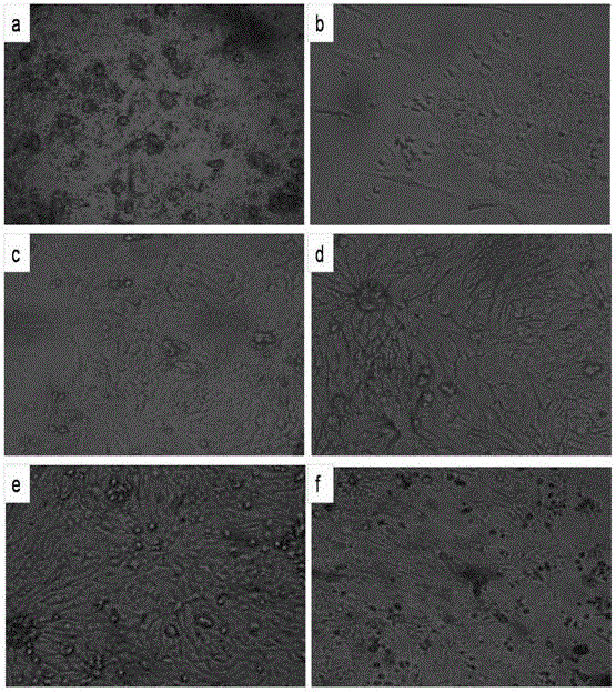
[0049] Aseptically separate tissue from the neck of the oviduct funnel near the follicle of a 26-week-old laying hen, cut the isolated tissue into pieces with a side length of 0.8 mm, and use 200 μg / mL penicillin and 200 μg / mL streptomycin. After washing the tissue with 7 mL of phosphate buffer solution to remove mucus, 0.25 wt% trypsin and 0.02 wt% EDTA were added to the tissue, and the tissue was digested in a constant temperature water bath at 37°C for 15 min. Then centrifuge at 670 rcf / min for 20 min, collect the precipitated cells, and then centrifuge the cell pellet at 670 rcf / min for 10 min. After the two centrifuges, use 10 mL of DMEM / F12 medium to suspend the precipitated tissue to obtain the suspended tissue, and use a cell sieve with a diameter of 100 μm to filter , collect the cells in the suspension tissue, centrifuge the collected cells at 670rcf / min for 10min, repeat the centrifugation twice, and then resuspend the cells with DMEM / F12 medium to obtain chicken ovi...

Embodiment 2

[0056] Aseptically isolate tissue from the neck of the oviduct funnel near the follicle of a 27-week-old laying hen, cut the isolated tissue into pieces with a side length of 0.9mm, and use penicillin 190U / mL, streptomycin 190U / mL After washing the tissue block with 5 mL of phosphate buffer solution to remove mucus, add 0.25 wt% trypsin and 0.02 wt% EDTA to the tissue, and digest the tissue in a constant temperature water bath at 37°C for 15 min. Then centrifuge at 670rcf / min for 20min, collect the precipitated cells, and then centrifuge the cell pellet at 670rcf / min for 10min.

[0057] The floating tissue was filtered with a cell sieve with a diameter of 100 μm, and the cells in the suspended tissue were collected. The collected cells were centrifuged at 670 rcf / min for 10 min, and the centrifugation was repeated twice, and then the cells were resuspended using DMEM / F12 medium to obtain chicken Fallopian tube epithelial cells. The resulting chicken oviduct epithelial cells w...

Embodiment 3

[0059] Aseptically separate tissue from the neck of the oviduct funnel near the follicle of a 25-week-old laying hen, cut the isolated tissue into pieces with a side length of 0.8mm, and use penicillin 210U / mL, streptomycin 210U / mL After washing the tissue block with 5 mL of phosphate buffer solution to remove mucus, add 0.25 wt% trypsin and 0.02 wt% EDTA to the tissue, and digest the tissue in a constant temperature water bath at 37°C for 15 min. Then centrifuge at 670 rcf / min for 20 min, collect the precipitated cells, and then centrifuge the cell pellet at 670 rcf / min for 10 min. After the two centrifuges, use 10 mL of DMEM / F12 medium to suspend the precipitated tissue to obtain the suspended tissue, and use a cell sieve with a diameter of 100 μm to filter , collect the cells in the suspension tissue, centrifuge the collected cells at 670rcf / min for 10min, repeat the centrifugation twice, and then resuspend the cells with DMEM / F12 medium to obtain chicken oviduct epithelial ...


PUM

PropertyMeasurementUnit
concentrationaaaaaaaaaa
diameteraaaaaaaaaa

Abstract

The invention provides a method for isolating and culturing primary chicken oviduct epithelial cells in vitro, comprising the following steps: 1) aseptically isolating the tissue from the infundibulum neck of the oviduct of laying hens close to the follicle; 2) using trypsin and EDTA to jointly digest the 3) Incubate the chicken oviduct epithelial cells at a temperature of 37° C. for 2 hours; 4) Perform cell culture on the cells incubated in step 3). The primary chicken fallopian tube epithelial cells isolated and cultured by the method have good effects, fast cell growth and good condition; the invention has short digestion time, good results, less miscellaneous cells, avoids the pollution of fibroblasts, and can obtain sound epithelial-like cells. cell population.

Description

technical field [0001] The invention relates to the field of animal cell culture, in particular to a method for separating and culturing chicken primary oviduct epithelial cells in vitro. Background technique [0002] In recent years, the decline in egg production of laying hens has become more and more serious. Chicken salpingitis and oviduct cysts are important reasons for the decline in egg production of various laying hens. The disease is caused by various causes of laying hens. It is characterized by blockage of fallopian tubes, caseous salpingitis, varying degrees of hydrosalpinx, and yolk peritonitis. Clinically, it is manifested as enlarged abdomen of sick chickens, decreased egg production, missing peak egg production, deformation of eggshells, increased feeding costs, and feed The low pay has brought great economic losses to the breeding industry, but its etiology and pathogenesis have puzzled people in the industry at home and abroad for decades. In order to reve...

Claims


Application Information

Patent Timeline
Patent Type & Authority Applications(China)
IPC IPC(8): C12N5/071
CPCC12N5/0682C12N5/0625C12N2500/32C12N2500/34C12N2501/11C12N2501/33
Inventor 杨霞李永涛王新卫刘红英陈陆赵军王川庆
Owner HENAN AGRICULTURAL UNIVERSITY
Why Eureka
Try Eureka