Ultra-wideband breast tumor imaging method based on magnetic resonance image compensation

A technology for magnetic resonance images and breast tumors, applied in image enhancement, image analysis, image data processing, etc., can solve problems such as rough interface and difficulty in accurately estimating the volume of glandular tissue

Inactive Publication Date: 2016-06-22
TIANJIN UNIV
View PDF3 Cites 5 Cited by
  • Summary
  • Abstract
  • Description
  • Claims
  • Application Information

AI Technical Summary

Problems solved by technology

Because the glandular tissue and adipose tissue in the breast are interwoven into a network structure, the interface between the two is very rough, and the volume of the glandular tissue is difficult to accurately estimate

Method used


Image

  • Ultra-wideband breast tumor imaging method based on magnetic resonance image compensation
  • Ultra-wideband breast tumor imaging method based on magnetic resonance image compensation
  • Ultra-wideband breast tumor imaging method based on magnetic resonance image compensation

Examples


Embodiment Construction

[0024] The present invention will be described below in conjunction with the accompanying drawings and embodiments.

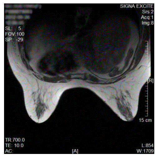
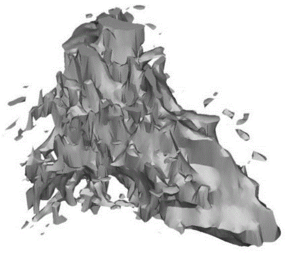
[0025] (1) figure 1 Shown are breast MRI slices. First, a discrete model of the gland is constructed based on the MRI slice atlas of the breast. After the MRI images are stacked at equal intervals, the gap between the slices is interpolated to construct a complete three-dimensional discrete breast model. Thresholds are defined for the degree value interval, and the interior of the breast is divided into various tissues, such as figure 2 shown. Directly extract the gland part in the discrete model, such as image 3 shown.

[0026] (2) Model the glandular structure so that the constructed model is similar to the original glandular structure. By comparing a large number of breast MRI images, the structure of the glandular tissue in the breast is approximated as an elliptical cone shape. This approximate shape can be compared with Good description of the gla...


PUM


Abstract

The invention relates to an ultra-wideband breast tumor imaging method based on magnetic resonance image compensation. The method comprises the following steps of based on a MRI slice atlas based on breasts, constructing a three-dimensional discrete breast model of a gland; modeling a gland structure and approximating structures of gland tissues in the breasts into an elliptic cone shape; using spatial coordinate transformation and ''righting'' an elliptic cone to an auxiliary coordinate system; creating an isosceles triangle area in a plane projection area of the elliptic cone, carrying out a finite element on the area and determining an elliptic cone surface capable of best describing a change trend of a rough gland surface; and carrying out microwave propagation time error estimation and accurate imaging. In the invention, ultra-wideband breast tumor imaging can be accurately performed.

Description

technical field [0001] The invention belongs to a breast tumor imaging method. Background technique [0002] Ultra-wideband (UWB) detection technology is developing rapidly in the field of non-destructive testing. UWB detection is widely used. This technology has broad application prospects in many fields such as ground penetrating radar, material flaw detection, wall-penetrating radar, breast tumor detection, etc. It has many advantages such as low cost of detection equipment, small size of equipment, and no radiation damage to the human body. This technology uses ultra-broadband microwaves to irradiate the object to be detected, and then relies on the principle of radar reflection to locate the target or image the target. In the application of ultra-broadband microwaves to the detection of human breast tumors, the complex structure of the breast leads to uneven and difficult to estimate the speed of ultra-fast microwave propagation, resulting in a decrease in imaging accu...

Claims


Application Information

Patent Timeline
Patent Type & Authority Applications(China)
IPC IPC(8): G06T7/00
CPCG06T7/0012G06T2207/10088G06T2207/30068
Inventor 肖夏王梁
Owner TIANJIN UNIV
Why Eureka
Try Eureka