Optical probe for endoscopic imaging

An optical probe and imaging technology, applied in the fields of endoscopy, application, medical science, etc., can solve the problem of incomplete cavity imaging, and achieve the effect of simplified structure, stable performance and easy assembly

Inactive Publication Date: 2016-07-20
SHANGHAI INST OF OPTICS & FINE MECHANICS CHINESE ACAD OF SCI
View PDF10 Cites 13 Cited by
  • Summary
  • Abstract
  • Description
  • Claims
  • Application Information

AI Technical Summary

Problems solved by technology

However, the disadvantage of this structure is that because the motor wire is looped back through the inner wall of the outer cannula from the tail, a part of the side wall of the lumen will be covered during scanning, resulting in incomplete imaging of the lumen by the OCT system.

Method used


Image

  • Optical probe for endoscopic imaging

Examples


Embodiment Construction

[0028] The technical solutions of the present invention will be further described below in conjunction with the accompanying drawings and through specific implementation methods.

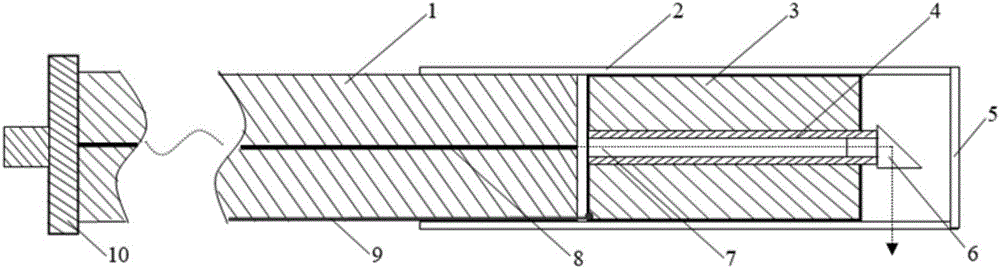
[0029] As shown in Figure 1, a non-imaging shadow optical probe based on a micro-motor includes an optical fiber connector 10, an optical fiber outer sleeve 1, a transparent outer sleeve 2, a micro-motor 3 assembled in the transparent outer sleeve, a self-focusing lens 7, and a right-angle prism 6. The rotating shaft 4 of the micro motor is hollow, and the self-focusing lens 7 is installed in it, and the central axis of the end of the optical fiber 8 in the optical fiber outer tube 1 is aligned with the central axis of the self-focusing lens 7 placed in the hollow rotating shaft 4 but There are fine intervals, the other end of the hollow rotating shaft 4 is glued to the right-angled edge 6, and the wire 9 of the micro-motor 3 is connected to the power supply from the side close to the end of the opti...


PUM


Abstract

An optical probe for endoscopic imaging comprises an optical fiber fixed in an outer optical fiber sleeve, a transparent outer sleeve as well as a micro-motor, a self-focusing lens and a rectangular prism which are assembled in the transparent outer sleeve, wherein one end of the outer optical fiber sleeve is embedded in the front end of the transparent outer sleeve, and the other end of the outer optical fiber sleeve is connected with an optical fiber connector; a rotary shaft of the micro-motor is hollow, and the self-focusing lens is mounted in the rotary shaft and close to the tail end of the optical fiber in the outer optical fiber sleeve; the axis of the optical fiber and the axis of the self-focusing lens keep on the same horizontal line and are separated; the rectangular prism is fixed at the tail end of the rotary shaft. With the adoption of the optical probe, 360-degree radial scanning of the vascular wall and other tissue can be realized, the problem of non-uniform rotational distortion due to the fact that a torque line is adopted to transmit torque to drive a probe to rotate is solved, and the problems of small radial scanning area of an optical probe provided with an MEMS (micro-electromechanical system) micro-mirror and image shadows caused by a wire in an optical probe provided with a micro-motor are solved.

Description

technical field [0001] The invention relates to the medical imaging instrument industry, in particular to an optical probe that can be used for endoscopic imaging. Background technique [0002] Optical coherence tomography (Optical Coherence Tomography, OCT) is an advanced imaging technology using the principle of weak coherent optical interferometer, which is widely used in the medical field. This technology puts forward strict requirements on the optical probe when performing endoscopic biomedical testing. The size of the probe should be as small as possible, and it should be stable during the working process and capable of 360-degree scanning. At present, many scientific research institutions at home and abroad have conducted related research on OCT optical probes for endoscopic imaging. [0003] A new type of non-radiation OCT probe and measurement system for blood vessel detection is disclosed in the invention patent literature of Chinese Patent Publication No. CN10488...

Claims


Application Information

Patent Timeline
Patent Type & Authority Applications(China)
IPC IPC(8): A61B1/05A61B5/00
CPCA61B1/05A61B1/00131A61B1/00165A61B5/0066A61B2576/00
Inventor 卢宇李中梁王向朝南楠贺琪欲陈艳王瑄潘柳华
Owner SHANGHAI INST OF OPTICS & FINE MECHANICS CHINESE ACAD OF SCI
Why Eureka
Try Eureka