Systems and methods for determining lesion depth by using fluorescence imaging

A technique of fluorescence intensity and depth, applied in the direction of using fluorescence emission for analysis, application, parts of surgical instruments, etc., can solve problems such as stagnation of success rate

Active Publication Date: 2016-10-12
GEORGE WASHINGTON UNIVERSITY +1
View PDF12 Cites 17 Cited by
  • Summary
  • Abstract
  • Description
  • Claims
  • Application Information

AI Technical Summary

Problems solved by technology

However, the success rate of AF ablation methods has remained relatively stagnant, with relapse rates estimated to be as high as 30% to 50% one year after the method

Method used


Image

  • Systems and methods for determining lesion depth by using fluorescence imaging
  • Systems and methods for determining lesion depth by using fluorescence imaging
  • Systems and methods for determining lesion depth by using fluorescence imaging

Examples


Embodiment

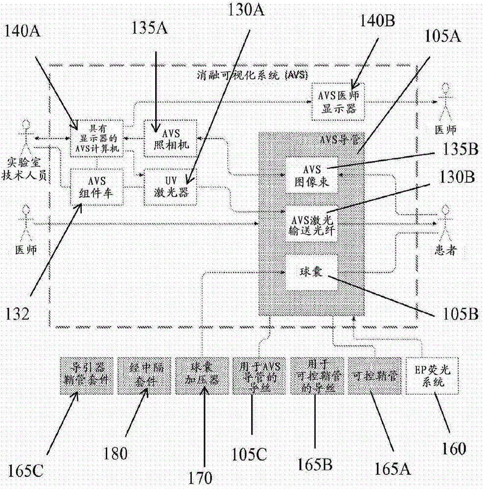
[0086] Experiments were performed using a functionally equivalent system to generate ablated lesions and lesion images to develop methods for lesion depth analysis. The experimental setup is described below.

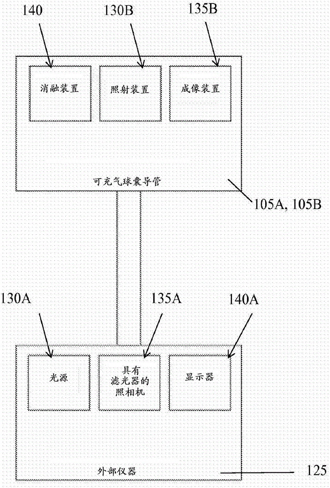
[0087] The NADH fluorescence system was provided to illuminate the epicardial surface using an LED spotlight (PLS-0365-030-07-S, Mightex Systems) with a peak wavelength of 365 nm. Emitted light was bandpass filtered at 460nm + / - 25nm and imaged using a CCD camera (Andor Ixon DV860) fitted with a low magnification lens. Fluorescence of NADH (fNADH) was imaged to monitor the status of epicardial tissue.

[0088] The RFA system provides RFA using a standard clinical RF generator (EPT 1000 Ablation System from Boston Scientific). The generator was electrically connected to the animal via a 4mm cooled Blazer ablation catheter (Boston Scientific) to deliver the lesion. Use grounding pads during ablation. The generator is set to temperature control mode. Cryoablation is pe...


PUM


Abstract

Systems, catheter and methods for treating Atrial Fibrillation (AF) are provided, which are configured to illuminate a heart tissue having a lesion site; obtain a mitochondrial nicotinamide adenine dinucleotide hydrogen (NADH) fluorescence intensity from the illuminated heart tissue along a first line across the lesion site; create a 2-dimensional (2D) map of the depth of the lesion site along the first line based on the NADH fluorescence intensity; and determine a depth of the lesion site at a selected point along the first line from the 2D map, wherein a lower NADH fluorescence intensity corresponds to a greater depth in the lesion site and a higher NADH fluorescence intensity corresponds to an unablated tissue. The process may be repeated to create a 3 dimensional map of the depth of the lesion.

Description

[0001] related application [0002] This application claims the benefit and priority of U.S. Application Serial No. 14 / 541,991, filed November 14, 2014, and claims the benefit and priority of U.S. Provisional Application Serial No. 61 / 904,018, filed November 14, 2013, The entire contents of both are incorporated herein by reference in their entirety. technical field [0003] The present disclosure generally relates to the medical procedure of applying ablative energy to the body to create a therapeutic lesion. In particular, the present disclosure relates to systems and methods for imaging lesions and tissue to determine lesion depth. Background technique [0004] Atrial fibrillation (AF) is the most common sustained cardiac arrhythmia worldwide, currently affecting millions of people. In the United States, AF is expected to affect 10 million people by 2050. AF is associated with increased mortality, morbidity, affects quality of life, and is an independent risk factor fo...

Claims


Application Information

Patent Timeline
Patent Type & Authority Applications(China)
IPC IPC(8): A61B5/00
CPCA61B5/0044A61B5/0071A61B5/0084A61B5/4836A61B18/02A61B18/12A61B2018/00351
Inventor 马可·A·梅卡德尔纳里内·萨尔瓦扬特伦斯·J·兰斯伯里肯尼斯·C·阿姆斯特朗奥马尔·阿米拉纳
Owner GEORGE WASHINGTON UNIVERSITY
Why Eureka
Try Eureka