Biological self-illumination three-dimensional imaging method based on pure optical system

A three-dimensional imaging and optical system technology, applied in image generation, analysis using fluorescence emission, medical science, etc., can solve problems such as adverse effects of reconstruction results, lack of anatomical information in three-dimensional homogeneous spatial information, etc., to improve secondary education Optical projection results, effective biological autofluorescence imaging, the effect of avoiding radiation hazards

Active Publication Date: 2016-11-09
INST OF AUTOMATION CHINESE ACAD OF SCI
View PDF4 Cites 12 Cited by
  • Summary
  • Abstract
  • Description
  • Claims
  • Application Information

AI Technical Summary

Problems solved by technology

[0005] In addition, because the three-dimensional homogeneous spatial information obtained by purely using optical projection surface reconstruction lacks anatomical information, replacing traditional CT imaging with optical projection surface reconstruction will adversely affect the reconstruction results

Method used


Image

  • Biological self-illumination three-dimensional imaging method based on pure optical system
  • Biological self-illumination three-dimensional imaging method based on pure optical system
  • Biological self-illumination three-dimensional imaging method based on pure optical system

Examples


Embodiment Construction

[0050] In order to make the object, technical solution and advantages of the present invention clearer, the present invention will be described in further detail below in conjunction with specific embodiments and with reference to the accompanying drawings.

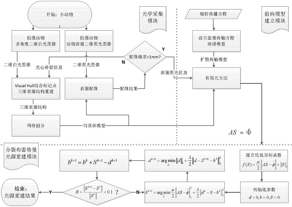
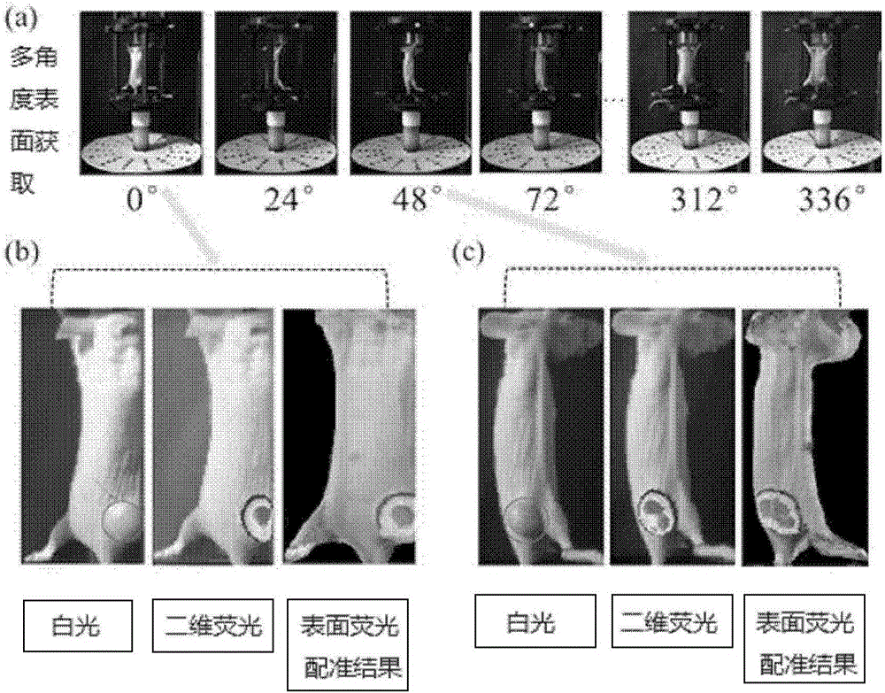
[0051] The invention is a pure optical biological self-luminescence three-dimensional imaging method combined with multi-angle optical projection surface reconstruction based on secondary correction and split Bregman reconstruction algorithm. Multi-angle optical projection surface reconstruction based on quadratic correction is a method to reconstruct the three-dimensional structure of the object surface with the help of the shooting results of multi-angle two-dimensional images, and provide spatial information for fluorescence registration, and use the fluorescence registration information to simultaneously The position deviation of a marker calculates the deviation of the optical center of the camera, and then corrects t...


PUM


Abstract

The invention discloses a biological self-illumination three-dimensional imaging method based on a pure optical system, comprising the steps of: obtaining the multi-angle two-dimensional white light image, two-dimensional fluoroscopic image and two-dimensional white light marking point distribution of a target object; reconstructing the three-dimensional surface structure of the target object; obtaining an isotropic body model through mesh generation; registering three-dimensional coordinates of a marker to obtain the distance deviation between a rotary central axis and an optical center outer normal vector; mapping two-dimensional fluoroscopic image fluorescence signals into the isotropic body model to obtain fluoroscopic surface light intensity information; building a photon diffusion transfer model in the target object; and establishing an optimal objective function and solving optimal intensity distribution in the target object, and furthermore obtaining biological autofluorescence luminescence light source distribution, i.e., biological autofluorescence imaging results. The biological self-illumination three-dimensional imaging method can be effectively applied to the whole body physiological metabolism research of the target object, has high reconstruction efficiency, and is suitable for cases with lower imaging system performance or poor experiment environment.

Description

technical field [0001] The invention belongs to the field of medical molecular imaging, and relates to computer vision and autofluorescence tomography methods, in particular to a 3D surface reconstruction that combines multi-angle optical images for optical projection and split Bregman-based autofluorescence tomography techniques. Background technique [0002] Optical imaging technology is an emerging molecular imaging technology in recent years. It has the advantages of high sensitivity, non-ionizing radiation, and easy operation. It has gradually become an important modality in the research of small animal in vivo imaging. A technology that realizes non-invasive real-time dynamic continuous imaging of biophysical processes inside biological tissues at the molecular level. It detects the distribution of the light source inside the target body through the acquired light intensity distribution on the surface of the target body, and can carry out quantitative research on the t...

Claims


Application Information

Patent Timeline
Patent Type & Authority Applications(China)
IPC IPC(8): G06T17/00G06T7/00A61B5/00
CPCA61B5/0071A61B5/0073G06T17/00G06T2200/08G06T2210/41
Inventor 田捷王坤高源张爽刘红波
Owner INST OF AUTOMATION CHINESE ACAD OF SCI
Why Eureka
Try Eureka