3D optical molecular imaging laparoscopic imaging system

A molecular imaging and imaging system technology, applied in the field of biomedical laparoscopy, can solve the problems of no surgeon, disconnection, and no 3D imaging, and achieve the effect of enriching depth information, improving accuracy, and precise fusion

Active Publication Date: 2016-12-21
SHANXI MEDICAL UNIV
View PDF6 Cites 25 Cited by
  • Summary
  • Abstract
  • Description
  • Claims
  • Application Information

AI Technical Summary

Problems solved by technology
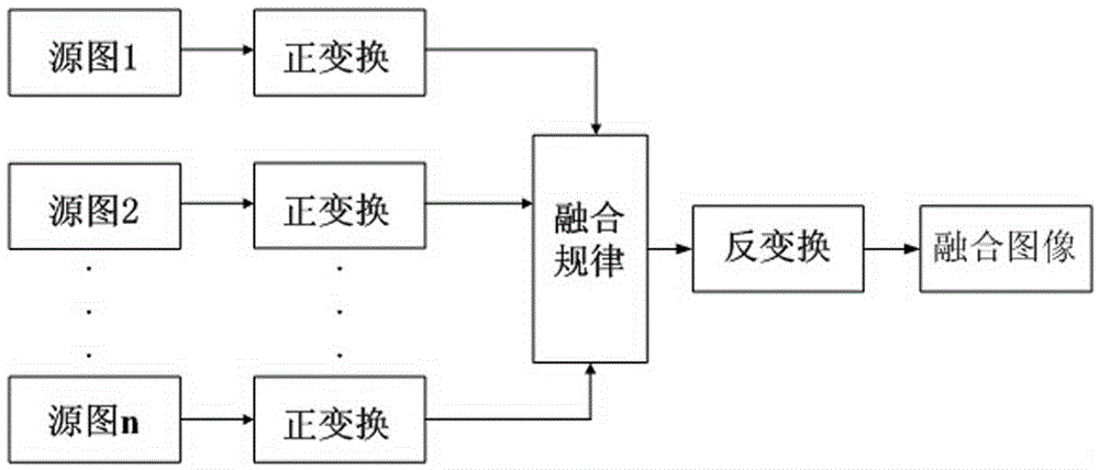
However, both the 2D images of traditional laparoscopy and the 3D images of 3D laparoscopy are images of anatomical structures, and neither have achieved precise functional images at the molecular level
[0007] In 2003, Professor John V. Frangioni of Harvard Medical School proposed the surgical operation of optical molecular image navigation

Method used


Image

  • 3D optical molecular imaging laparoscopic imaging system
  • 3D optical molecular imaging laparoscopic imaging system
  • 3D optical molecular imaging laparoscopic imaging system

Examples


Embodiment

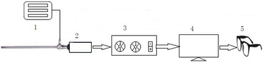
[0056] 1. CCD selection

[0057] The color CCD is 2 / 3, high-performance CCD, progressive scanning to capture moving images, super high resolution 1200TVL, 1920×1080P full HD output digital image, working voltage is 12V; black and white CCD is 1 / 2 inch, HAD technology, high Sensitivity CCD, near-infrared high resolution 752×582 pixels. CCD is an image sensor, also called an image controller

[0058] 2. Prism selection

[0059] The prism divides the light output from the lens body into visible light and near-infrared light. The ICG fluorescent dye is commonly used. The excitation light of ICG is 780±10nm, the emitted light is 845±10nm, and the wavelength of visible light is 400-700nm, so the spectroscopic prism The function is to divide the output light into two beams of 400-700nm and 845±10nm, and filter out the laser light of 780±10nm.

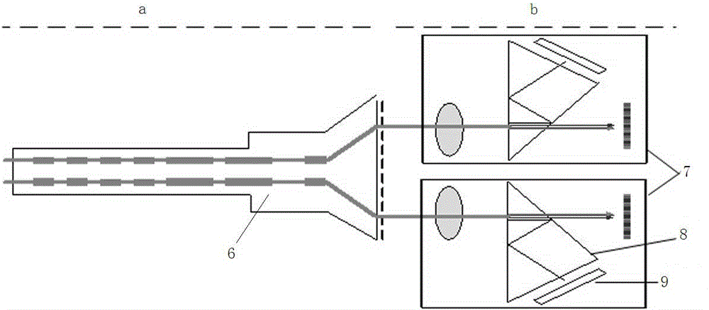
[0060] 3. The optical path design of a single set of dual-channel 2CCD

[0061] The light output by the laparoscope is divided into visib...


PUM


Abstract

The invention belongs to the technical field of biomedical laparoscope, and particularly relates to a 3D optical molecular imaging laparoscopic imaging system, aiming at solving the problem in existing laparoscope technology and fluorescent laparoscopes. The system comprises a 3D laparoscope, wherein two output optical channels of the 3D laparoscope are parallel; a dual-channel charge coupled device (CCD) is respectively arranged outside the 3D laparoscope corresponding to two eye lenses, and a beam splitter prism is arranged in each dual-channel CCD; a cut-off filter is arranged at the near infrared channel behind the beam splitter prism, and a black-white near infrared CCD is arranged behind the cut-off filter; and a colored CCD is arranged at the visible light channel behind the beam splitter prism. According to the 3D optical molecular imaging laparoscopic imaging system, a target optical molecular image is precisely integrated with a visible light image, and is displayed in a 3D manner, so that the 3D structure of a target focus and the 3D anatomical relationship of the 3D structure and surrounding tissues can be displayed precisely, rich deep information is provided to doctors, and the accuracy of doctor operation and scientific researches can be greatly improved.

Description

technical field [0001] The invention belongs to the technical field of biomedical laparoscopy, and in particular relates to a 3D optical molecular image laparoscopy imaging system. Background technique [0002] With the development of modern science and technology and medical equipment, medical surgery will develop from minimally invasive surgery to precision surgery and molecular targeted surgery, and the accuracy of surgery will also develop from organs, tissues, and cells to the molecular level. Diagnosis and treatment will become an important direction of future medical development. [0003] Molecular imaging is a science that uses imaging technology to qualitatively and quantitatively study molecular changes and their biological behavior in vivo by displaying the molecular dynamics characteristics of specific molecules at the tissue, cellular and subcellular levels. At present, medical imaging includes: X-ray, CT, MR, and ultrasound imaging that show the anatomical str...

Claims


Application Information

Patent Timeline
IPC IPC(8): A61B1/313A61B1/04A61B1/06A61B1/00A61B5/00
CPCA61B1/00009A61B1/00131A61B1/00197A61B1/043A61B1/0661A61B1/3132A61B5/0071A61B5/0075A61B5/0086A61B5/7235A61B5/7253
Inventor 杨晓峰
Owner SHANXI MEDICAL UNIV
Why Eureka
Try Eureka