Ultrasonic wide-view imaging method for spinal scoliosis

A wide-field imaging and scoliosis technology, applied in the field of medical ultrasound wide-field imaging, can solve the problems of difficulty in accurately obtaining accurate imaging results of the spine, difficulty in ensuring the accuracy of measurement results, and difficulty in calculating the angle of scoliosis. Flexibility and convenience, easy control, simple method effects

Inactive Publication Date: 2017-02-01
SOUTH CHINA UNIV OF TECH
View PDF6 Cites 21 Cited by
  • Summary
  • Abstract
  • Description
  • Claims
  • Application Information

AI Technical Summary

Problems solved by technology

[0004] Traditional ultrasound imaging of scoliosis uses a probe to scan directly above the spine, but because the vertebrae of the spine are densely spaced and too close to the skin, it is difficult to distinguish each vertebra from the ultrasound image. In addition, the probe is used to scan directly above the spine , the scanning direction of the ultrasound probe is difficult to control, and it is difficult to obtain accurate imaging results of the spine, so it is difficult to calculate the angle of scoliosis
And when the above method is used for scanning, there is a one-time measurement error, and it is difficult to ensure the accuracy of the measurement results

Method used


Image

  • Ultrasonic wide-view imaging method for spinal scoliosis
  • Ultrasonic wide-view imaging method for spinal scoliosis

Examples


Embodiment

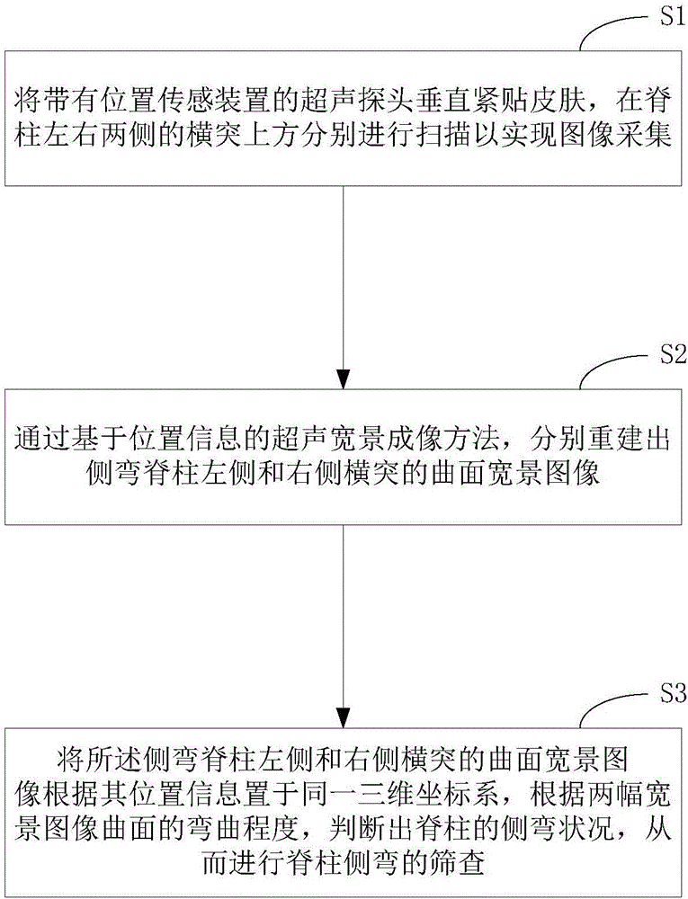
[0022] Such as figure 1 as shown, figure 1 A flow chart of an ultrasonic wide-view imaging method for scoliosis is disclosed. In this embodiment, an ultrasonic wide-view imaging method for scoliosis specifically includes the following steps:

[0023] S1. Put the ultrasound probe with the position sensing device close to the skin vertically, and scan above the transverse processes on the left and right sides of the spine to realize image acquisition;

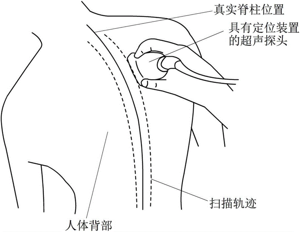
[0024] In a specific application, a positioning device is installed on the ultrasound probe to record the position information of the probe movement, and the probe bound with the positioning device is closely attached to the skin of the spine, and scanning is performed on the transverse processes on the left and right sides of the spine respectively, as shown in Fig. figure 2 As shown by the two dotted line trajectories in the image, at the same time, it is ensured that the person being scanned remains as still as possible duri...


PUM


Abstract

The invention discloses an ultrasonic wide-view imaging method for spinal scoliosis, which comprises the following steps: S1, enabling an ultrasonic probe with a position sensing device to vertically and closely stick to skins, respectively scanning upper parts of transverse processes on the left and right sides of a spine to realize image acquisition; S2, respectively reestablishing curved surface wide-view images of transverse processes on the left and right sides of a scoliosis spine through a position information-based ultrasonic wide-view imaging method; S3, placing the curved surface wide-view images of the transverse processes on the left and right sides of the scoliosis spine in the same three-dimensional coordinate system according to the position information, judging the scoliosis status of the spine according to the bending degree of curved surfaces of the two wide-view images, and screening spinal scoliosis. According to the ultrasonic wide-view imaging method, the scanning flexibility and the scanning convenience of the ultrasonic probe are improved, the ultrasonic probe is easy to control and different vertebrae are distinguished, the accuracy of an imaging result is enhanced through scanning measurement twice, and the method has the characteristics of simplicity, convenience for operation and high practicability.

Description

technical field [0001] The invention relates to medical ultrasonic wide-view imaging technology, in particular to an ultrasonic wide-view imaging method for scoliosis. Background technique [0002] Under normal circumstances, the human spine grows symmetrically about the trunk. When the spine bends to one side at an angle of more than 10°, it is called scoliosis. When the scoliosis is severe, it will seriously endanger the normal growth and development of the patient, and surgical correction is required. The key to timely treatment of scoliosis is early detection. [0003] At present, the screening method for scoliosis mainly uses standing X-ray film, which is more accurate, but due to the strong radiation of X-ray, it is chosen to use weak radiation and relatively cheap ultrasound imaging to scan the side of the spine. It has good practical value and significance to screen the bending situation. [0004] Traditional ultrasound imaging of scoliosis uses a probe to scan di...

Claims


Application Information

Patent Timeline
Patent Type & Authority Applications(China)
IPC IPC(8): A61B8/08
CPCA61B8/0833A61B8/0875A61B8/4444A61B8/52
Inventor 黄庆华曾昭峥赵夏黄仙海
Owner SOUTH CHINA UNIV OF TECH
Why Eureka
Try Eureka